Submitted Successfully!
To reward your contribution, here is a gift for you: A free trial for our video production service.
Thank you for your contribution! You can also upload a video entry or images related to this topic.
Version Summary Created by Modification Content Size Created at Operation
1 + 2765 word(s) 2765 2021-08-18 08:29:29 |
2 format change Meta information modification 2765 2021-08-19 11:30:17 |

Video Upload Options

Do you have a full video?

Confirm

Are you sure to Delete?
Cite
If you have any further questions, please contact Encyclopedia Editorial Office.
Ehrmann, A.; Tanzli, E. Electrospun Nanofibrous Membrane. Encyclopedia. Available online: https://encyclopedia.pub/entry/13322 (accessed on 24 April 2024).
Ehrmann A, Tanzli E. Electrospun Nanofibrous Membrane. Encyclopedia. Available at: https://encyclopedia.pub/entry/13322. Accessed April 24, 2024.
Ehrmann, Andrea, Ewin Tanzli. "Electrospun Nanofibrous Membrane" Encyclopedia, https://encyclopedia.pub/entry/13322 (accessed April 24, 2024).
Ehrmann, A., & Tanzli, E. (2021, August 18). Electrospun Nanofibrous Membrane. In Encyclopedia. https://encyclopedia.pub/entry/13322
Ehrmann, Andrea and Ewin Tanzli. "Electrospun Nanofibrous Membrane." Encyclopedia. Web. 18 August, 2021.
Electrospun Nanofibrous Membrane
Edit

In biotechnology, the field of cell cultivation is highly relevant. Cultivated cells can be used, for example, for the development of biopharmaceuticals and in tissue engineering. Commonly, mammalian cells are grown in bioreactors, T-flasks, well plates, etc., without a specific substrate. Nanofibrous mats, however, have been reported to promote cell growth, adhesion, and proliferation. Here, we give an overview of the different attempts at cultivating mammalian cells on electrospun nanofiber mats for biotechnological and biomedical purposes. Starting with a brief overview of the different electrospinning methods, resulting in random or defined fiber orientations in the nanofiber mats, we describe the typical materials used in cell growth applications in biotechnology and tissue engineering. The influence of using different surface morphologies and polymers or polymer blends on the possible application of such nanofiber mats for tissue engineering and other biotechnological applications is discussed. Polymer blends, in particular, can often be used to reach the required combination of mechanical and biological properties, making such nanofiber mats highly suitable for tissue engineering and other biotechnological or biomedical cell growth applications.

cell growth electrospinning

1. Introduction

The cultivation of cells in T-flasks is a common method in biotechnology. For the classical cultivation of adherent cells in roller bottles or cell culture pellets, manufacturing costs are very high [1]. Microcarriers and hollow fibers are an alternative to the classical systems and can replace these bioreactor systems [2][3]. Such hollow fibers can be prepared from various polymers [4][5].
Today’s single-use fixed-bed bioreactors provide consistent product quality from batch to batch, which is vital in pharmaceutical processes. With continuous bioprocesses, smaller production volumes and higher product concentrations are possible. Such ready-to-use bioreactors are packed with non-woven macroporous carriers or rolled membranes. The fabric often consists of polyethylene terephthalate (PET) microfibers, which are surface treated to make them hydrophilic and biocompatible for cell attachment [6][7][8].
There are, however, approaches to support cell growth more effectively, e.g., by using nanofiber mats as substrates for cell growth [9]. Such new nanostructured substrates can improve cell growth in tissue engineering and biotechnology by guiding cell growth and even improving stem cell differentiation. On the other hand, tissue engineering often necessitates 3D substrates, which cannot be reached by bioreactors or T-flasks, while nanofiber mats can be draped according to the requirements of an experiment.
Such nanofiber mats can be produced by electrospinning from various polymers and polymer blends, as well as polymers with nanoparticles included [10][11][12]. With electrospinning techniques being developed further, nanofiber mats have been increasingly used for biotechnological applications, including cell cultivation [13][14][15][16].
Besides the large specific surface of such nanofiber mats, specific electrospinning techniques or substrate preparations enable spinning oriented fibers, which is not only supportive in terms of mechanical properties, but also for growing cells on these substrates in a defined orientation and in many cases with a higher proliferation rate [17][18][19][20].
Here, we give a brief overview of recent electrospinning techniques, followed by reviewing materials typically used for the preparation of electrospun nanofiber mats as scaffolds for mammalian cell growth. The focus is put on Chinese hamster ovary (CHO) cells, which are often used for experiments in such cell cultures, not only for the production of therapeutic proteins, but also as model cells for biotechnological studies [21][22][23].

2. Gelatin and Collagen

Collagen belongs to the native extracellular matrix (ECM) of the human body. The ECM is responsible for providing the mechanical structure of tissues, as well as for wound healing and controlling other cellular processes [24]. Thus, collagen belongs to the highly interesting materials for tissue engineering. Gelatin, which is derived from collagen by partial hydrolysis, has partly similar and partly different physical properties, varying depending on the origin and processing of the material [25][26].
As usual for many biopolymers, collagen and gelatin are water-soluble and thus need a crosslinking step [27]. Alternatively, the water-resistance and mechanical properties can be enhanced by blending them with water-stable polymers. This is why many studies found in the literature report on blends of collagen or gelatin with other polymers.
For example, Chen et al. prepared polyurethane/collagen core-shell nanofibers by coaxial electrospinning that were crosslinked by glutaraldehyde (GA) vapor [28]. Thermoplastic polyurethanes (TPUs) are known to have not only good mechanical properties, but also good biocompatibility, resulting in their frequent use in implants, catheters, etc. [29]. Special medical-grade TPU can additionally avoid in vivo degradation [30]. The coaxial electrospinning technique used here allows spinning two different polymers through the inner and the outer part of the needle, driven by two independent syringe pumps and fed from separate reservoirs [28]. In comparison with pure collagen, the core-shell fibers showed significantly improved mechanical properties, combined with the highest viability of pig iliac endothelial cells (PIECs) cultured on the different electrospun nanofiber mats, even higher than on pure collagen fibers. All nanofiber mats showed a better cell viability than reference tests on coverslips. The authors mentioned that the fiber diameters, mat porosity, and mechanical properties of the nanofiber mats significantly influenced cell growth and migration, which resulted in compound nanofibers—especially produced by coaxial electrospinning of collagen around TPU—having the best growth conditions. They also mentioned that the coaxially collagen-coated TPU fibers were much more attached cells, while simply coating a pure TPU nanofiber mat macroscopically with collagen did not have the same effect of 3D ingrowth into the scaffold, showing the importance of combining material and structure. Similar results were found by the same group with TPU/collagen blended nanofibers [31].
A polymer blend from collagen and poly((D,L-lactic)-co-glycolide) (PLGA) was investigated by Yang et al. [32]. PLGA belongs to the synthetic biodegradable polymers, which are often studied for tissue engineering applications, but usually show insufficient cell adhesion and proliferation if used purely [33]. Blending PLGA with collagen, however, led to electrospun nanofiber mats which did not need to be crosslinked to avoid swelling in culture medium and on which human dermal fibroblasts (HDFs) and mice fibroblasts containing green fluorescent protein (GFP) showed a significantly higher viability than on pure PLGA nanofiber mats [32]. This was attributed to the chemical composition of the matrix material, since the fiber diameters were kept constant in all nanofiber mats. The authors explained that collagen and its integrin-binding domains facilitated the cell attachment for anchorage-dependent cell types as the fibroblasts used in this study. A comparison with tissue culture plates additionally showed much higher cell numbers inside the nanofibrous mats due to the available void volume.
Quite a different approach was chosen by Akhshabi et al., who combined collagen with chondroitin sulfate, a negatively sulfated glycosaminoglycan [34]. The electrospun collagen/chondroitin nanofiber mat was cross-linked with EDC/NHS (1-ethyl-3-(3-dimethyl-aminopropyl)-1-carbodiimide hydrochloride/N-hydroxy succinimide), before cytocompatibility was investigated using corneal epithelial cells. Crosslinking was found to significantly increase the biostability of the nanofiber mats, while chondroitin was reported to significantly promote cell proliferation, as compared to pure collagen. This was explained by the role of chondroitin sulfate, which induces skin tissue regeneration [35], osteoblast adhesion [36], and fibroblast proliferation [37].
Due to the chemical similarity of gelatin and collagen, gelatin can be expected to show similar effects in increasing cell adhesion and proliferation. Baiguera et al. used genipin cross-linked gelatin nanofiber mats as the base to include decellularized rat brain extracellular matrix as an active agent, before mesenchymal stromal cells (MSCs) from rat bone marrow were seeded onto these scaffolds [38]. They found the MSCs grew not only on top of the substrate, but also in the inner volume of the decellularized samples. Seeding these cells on pure gelatin nanofiber mats and those with an additional decellularized rat brain extracellular matrix showed growth on both sides of the nanofiber mats in multilayered cultures, especially for the second tissue. Incorporation of a decellularized rat brain extracellular matrix into gelatin nanofiber mats, however, was found to trigger differentiation of MSCs towards neural/glial precursor cells.
An interesting approach was chosen by Li et al., who blended gelatin with the conductive polymer polyaniline (PAni) to prepare conductive electrospun scaffolds [39]. With conductive scaffolds, cell attachment, proliferation, migration, and even differentiation can be stimulated [40]. PAni, on the other hand, is known to be biocompatible [41] and thus a good candidate to make a nanofiber mat conductive. Li et al. seeded rat cardiac myoblast cells on these electrospun nanofiber mats and found they supported cell attachment and proliferation with different amounts of PAni, as compared to glass and other substrates, but also in comparison with pure gelatin nanofiber mats 
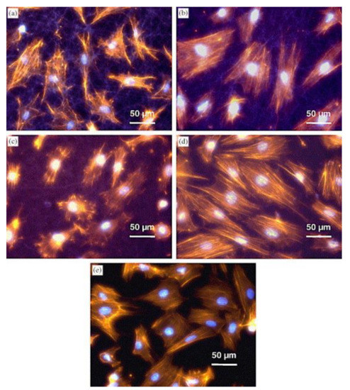
[39]. They found the cell morphology depended on the substrate structure, with cells growing on fibers with larger diameters showing microfilament-rich pseudopodia attached to single fibers (Figure 1a–c), while cells on thinner fibers were more spread out and exhibited a smooth muscle-like morphology (Figure 1d), similar to cells grown on glass substrates (Figure 1e) [39]. This was attributed to the higher roughness of the fibrous substrates, which provided a larger surface area on which cells could attach, as compared to smooth glass substrates and tissue culture-treated polystyrene, which is commonly regarded as the gold-standard.
Figure 1. Morphology of myoblast cells 20 h after seeding on: (a) gelatin nanofiber mat; (b) 15:85 PAni-gelatin blend nanofibers; (c) 30:70 PAni-gelatin blend nanofibers; (d) 45:55 PAni-gelatin blend nanofibers; and (e) glass matrices. Staining for nuclei-bisbenzimide and actin cytoskeleton-phalloidin. Reprinted from [39], with permission from Elsevier.
Like collagen, gelatin can also be blended with water-resistant or slowly degrading polymers to reduce or avoid crosslinking. Gautam et al. report about PCL/gelatin blended nanofiber mats used for tissue engineering [42]. They seeded mouse fibroblast cells on nanofiber mats with different blend ratios and found significantly increased cell viability and cell proliferation for the blends as compared to pure PCL scaffolds. This was attributed to the presence of gelatin in these nanofiber mats, which is generally known to result in good cell adhesion and proliferation. Combinations of gelatin with PCL are thus often found in the literature [43][44][45].
There are, however, many other gelatin blends reported to be advantageous. Comparing PLGA and PLGA/gelatin blended nanofiber mats, Meng et al. found increased cell viability on PLGA/gelatin blends compared to pure PLGA, and in addition they also found better MC3T3-E1 cell proliferation and guidance for aligned nanofibers than for randomly oriented ones [46]. They mentioned that avoiding crosslinking induced easier cell attachment to the scaffolds, as crosslinking decreased bioactivity and hydrophilicity of the nanofiber mats. Ba Linh et al. investigated polyvinyl alcohol (PVA)/gelatin nanofiber mats after physical crosslinking by methanol and reported osteoblasts to attach firmly on these scaffolds, facilitated by gelatin and the crosslinking process [47]. Moreover, the hydrophilic nature of PVA and gelatin, as well as the interconnected porous structure of the nanofiber mats, were found to be advantageous for cell proliferation and multilayer growth. A special polyurethane “Tecophilic” was chosen by Vatankhah et al. as a blend partner for gelatin to produce tubular scaffolds for blood vessels, which showed good mechanical properties, higher cell viability and proliferation than pure polyurethane, and good compatibility with blood [48]. Here again, a deep cellular infiltration into the nanofibrous scaffold was found, which would be impossible on the common flat surfaces, allowing for a larger number of attached cells per area. In addition, triple-blends including gelatin were tested for tissue engineering applications [49][50][51].

3. Chitosan and Chitin

Chitin belongs to the highly abundant natural polymers and is mainly found in marine crustaceans, shrimps, and crabs. It is insoluble in common solvents. Chitosan, on the other hand, is an important derivative of chitin, obtained by partial deacetylation of chitin under alkaline conditions or by enzymatic hydrolysis and is soluble in acidic aqueous media [52].
In spite of the problems in dissolving chitin, it is regularly reported in the literature as a possible substrate material for tissue engineering [53], since it shows similar properties to glycosaminoglycan, which is part of the ECM [54]. Noh et al. thus prepared electrospun chitin nanofiber mats [54]. To dissolve the chitin powder, it was first irradiated by Co60 γ-irradiation and then dissolved in 1,1,1,3,3,3-hexafluoro-2-propanol (HFIP) for 20 days. Electrospinning resulted in ultrafine nanofibers with an average diameter 163 nm. These nanofiber mats degraded in vitro faster than chitin microfibers, promoted cell attachment and spreading of normal human keratinocytes and fibroblasts better than the chitin microfibers, and after coating with type I collagen also promoted cellular response in all cells under investigation. The authors interpreted these findings as a higher functional activity of collagen-coated chitin nanofibers in terms of cell attachment and spreading for normal human keratinocytes as well as fibroblasts. In a similar manner, Min et al. prepared a chitin solution by γ-irradiation and dissolving the resulting chitin in HFIP [55]. In addition, they investigated deacetylation for transforming the original chitin matrix into a chitosan matrix.
Most groups, however, combine chitin or its derivatives with other polymers. For example, Shalumon et al. blended the water-soluble carboxymethyl chitin with PVA to prepare an aqueous electrospinning solution 
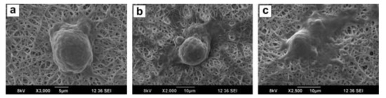
[56]. The resulting nanofiber mats were cross-linked by glutaraldehyde vapor before human mesenchymal stem cells (hMSCs) were seeded on them. The cells were found to attach and spread in the washed nanofiber mats, which supported cell adhesion and proliferation (Figure 2).
Figure 2. SEM images of hMSCs attached on the surfaces of CMC/PVA scaffolds after (a) 12 h; (b) 24 h; and (c) 48 h of incubation. Reprinted from [56], with permission from Elsevier.
Pangon et al. prepared chitosan/PVA nanofibers with chitin whiskers in different concentrations from succinic acid/water as solvent [57]. The resulting nanofiber mats were crosslinked by GA vapor, followed by mineralizing hydroxyapatite onto them by applying ten-fold concentrated simulated body fluid and seeding MC3T3-E1 osteoblast cells. They found an increased cell viability and proliferation due to the addition of the chitin whiskers, as compared to pure chitosan/PVA nanofiber mats. This effect was attributed to the clearly modified surface of the nanofiber mats, which showed a rough, uneven structure due to the hybridization with hydroxyapatite, and resulting in significantly improved cell viability and proliferation.
Electrospun nanocomposites of chitin nanofibrils and PCL were prepared by Ji et al. [58]. The chitin nanofibrils were dispersed in 2,2,2-trifluoroethanol (TFE) in which PCL could also be dissolved, allowing for spinning nanofibers with different chitin:PCL ratios, which showed strongly decreasing average diameters with increasing chitin content. For high chitin nanofibril contents, the water contact angle was significantly reduced. When seeding human dermal fibroblasts (hDF) on these nanofiber mats, the nanofiber mats with high chitin content, in particular, showed cell penetration and migration inside the scaffold; oppositely to pure PCL nanofiber mats, where the cells stayed on the surface. This finding is similar to those reported in other papers, where 3D structures and increased surface areas generally supported cell growth due to the improved possibility of cell infiltration and migration into the scaffold. The authors mentioned that for this effect, the pores in the electrospun nanofiber mats do not have to be larger than the cell diameters, but cells were found to perform amoeboid movements to migrate even through smaller pores by pushing the neighboring fibers away. Instead, hydrophilicity and biochemical signals were found to be more important to promote cell ingrowth than perfectly fitting pore sizes.
Min et al. used an electrospinning method based on two separate syringes to prepare PLGA nanofibers with chitin electrosprayed nanoparticles [59]. They seeded normal human oral keratinocytes and normal human epidermal keratinocytes on these nanofiber composites and found the PLGA/chitin nano-composite fibers to be well suitable for tissue engineering scaffolds, due to their biomimetic 3D structure, similar to the collagen/glycosaminoglycan composite structure in the extracellular matrix.
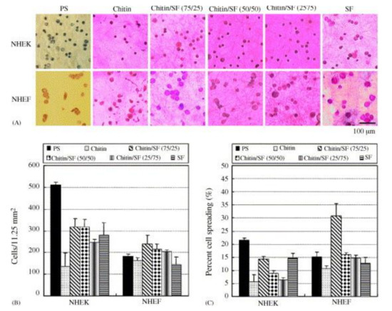
Another blend was investigated by Park et al., who used electrospinning to prepare chitin/silk fibroin (SF) blends from HFIP as solvent [60]. Normal human epidermal keratinocytes (NHEK) and fibroblasts (NHEF) were seeded onto these nanofiber mats after crystallization of the SF fraction in water vapor. While for attachment of NHEK cells, the reference polystyrene substrate performed best, cell spreading of NHEF cells was superior on chitin/SF nanofiber mats, as visible in Figure 3. This finding was explained by the high biocompatibility and other supportive properties of chitin and SF.
Figure 3. (A) Photographs and (B) numbers of NHEK and NHEK adhered on the substrates; (C) percentage of cell spreading (n = 4). Reprinted from [60], with permission from Elsevier.
As mentioned before, chitosan combines many of the positive physical and chemical properties of chitin with water-solubility, making it more easily electrospinnable. This is why diverse groups investigated electrospun chitosan or chitosan blend fibers. A recent review of electrospinning chitosan-based solutions for tissue engineering was given by Qasim et al. [61]. Some exemplary blend partners for chitosan are poly(ethylene oxide) PEO [62][63], collagen [64], PVA [65][66], PCL [67], and hydroxyapatite [68].
After this brief overview of materials correlated to the extracellular matrix, the next sections - which can be found in the original paper - will show progress in using electrospun man-made polymers as scaffolds in tissue engineering.

References

  1. Chatel, A. A brief history of adherent cell culture: Where we come from and where we should go. BioProcess Int. 2019, 17, 44–49.
  2. Whitford, W.G.; Hardy, J.C.; Cadwell, J.J.S. Single-use, continuous processing of primary stem cells. BioProcess Int. 2014, 12, 26–32.
  3. Simon, M. Bioreactor design for adherent cell culture. The bolt-on bioreactor project, part 1: Volumetric productivity. BioProcess Int. 2015, 13, 28–33.
  4. Allan, S.J.; De Bank, P.A.; Ellis, M.J. Bioprocess design considerations for cultured meat production with a focus on the expansion bioreactor. Front. Sustain. Food Syst. 2019, 3, 44.
  5. GE Healthcare Life Sciences. Microcarrier Cell Culture, Principles and Methods. 2013. Available online: http://www.gelifesciences.co.kr/wp-content/uploads/2016/07/023.8_Microcarrier-Cell-Culture.pdf (accessed on 30 June 2021).
  6. Lennaertz, A.; Knowles, S.; Drugmand, J.C.; Castillo, J. Viral vector production in the integrity iCELLis single-use fixed-bed bioreactor, from bench-scale to industrial scale. BMC Proc. 2013, 7, P59.
  7. Dohogne, Y.; Collignon, F.; Drugmand, J.C. Scale-X Bioreactor for Viral Vector Production. Proof of Concept for Scalable HEK293 Cell Growth and Adenovirus Production, Univercell Application Note. 2019. Available online: https://www.univercells.com/app/uploads/2019/05/scale-X%E2%84%A2-bioreactor-for-viral-production-Adeno_SFM.pdf (accessed on 30 June 2021).
  8. Drugmand, J.C.; Aghatos, S.; Schneider, Y.J. Growth of mammalian and lepidopteram cells on BioNOC® II disks, a novel macroporous microcarrier. In Cell Technology for Cell Products; Smith, R., Ed.; Springer: Heidelberg, Germany, 2007; pp. 781–784.
  9. Wehlage, D.; Blattner, H.; Sabantina, L.; Böttjer, B.; Grothe, T.; Rattenholl, A.; Gudermann, F.; Lütkemeyer, D.; Ehrmann, A. Sterilization of PAN/gelatine nanofibrous mats for cell growth. Tekstilec 2019, 62, 78–88.
  10. Teo, W.E.; Ramakrishna, S. A review on electrospinning design and nanofibre assemblies. Nanotechnology 2006, 17, R89.
  11. Ramakrishna, S.; Fujihara, K.; Teo, W.E.; Yong, T.; Ma, Z.; Ramaseshan, R. Electrospun nanofibers: Solving global issues. Mater. Today 2006, 9, 40–50.
  12. Grothe, T.; Wehlage, D.; Böhm, T.; Remche, A.; Ehrmann, A. Needleless electrospinning of PAN nanofibre mats. Tekstilec 2017, 60, 290–295.
  13. Gao, S.T.; Tang, G.S.; Hua, D.W.; Xiong, R.H.; Han, J.Q.; Jiang, S.H.; Zhang, Q.L.; Huang, C.B. Stimuli-responsive bio-based polymeric systems and their applications. J. Mater. Chem. B 2019, 7, 709–729.
  14. Sabantina, L.; Kinzel, F.; Hauser, T.; Többer, A.; Klöcker, M.; Döpke, C.; Böttjer, R.; Wehlage, D.; Rattenholl, A.; Ehrmann, A. Comparative study of Pleurotus ostreatus mushroom grown on modified PAN nanofiber mats. Nanomaterials 2019, 9, 475.
  15. Klinkhammer, K.; Seiler, N.; Grafahrend, D.; Gerardo-Nava, J.; Mey, J.; Brook, G.A.; Möller, M.; Dalton, P.D.; Klee, D. Deposition of electrospun fibers on reactive substrates for In Vitro investigations. Tissue Eng. Part C 2009, 15, 77–85.
  16. Mamun, A. Review of possible applications of nanofibrous mats for wound dressings. Tekstilec 2019, 62, 89–100.
  17. Bertocchi, M.J.; Simbana, R.A.; Wynne, L.H.; Lundin, J.G. Electrospinning of tough and elastic liquid crystalline polymer-polyurethane composite fibers: Mechanical properties and fiber alignment. Macromol. Mater. Eng. 2019, 304, 1900186.
  18. Bazrafshan, Z.; Stylios, G.K. A novel approach to enhance the spinnability of collagen fibers by graft polymerization. Mater. Sci. Eng. 2019, 94, 108–116.
  19. Guarino, V.; Iannotti, V.; Ausanio, G.; Ambrosio, L.; Lanotte, L. Elastomagnetic nanofiber wires by magnetic field assisted electrospinning. Express Polym. Lett. 2019, 13, 419–428.
  20. Johnson, C.D.L.; Ganguly, D.; Zuidema, J.M.; Cardina, T.J.; Ziemba, A.M.; Kearns, K.R.; McCarthy, S.M.; Thompson, D.M.; Ramanath, G.; Borca-Tasciuc, D.A.; et al. Injectable, magnetically orienting electrospun fiber conduits for neuron guidance. ACS Appl. Mater. Interfaces 2019, 11, 356–372.
  21. Möller, J.; Korte, K.; Pörtner, R.; Zeng, A.P.; Jandt, U. Model-based identification of cell-cycle-dependent metabolism and putative autocrine effects in antibody producing CHO cell culture. Biotechnol. Bioeng. 2018, 115, 2996–3008.
  22. Wippermann, A.; Rupp, O.; Brinkrolf, K.; Hoffrogge, R.; Noll, T. The DNA methylation landscape of Chinese hamster ovary (CHO) DP-12 cells. J. Biotechnol. 2015, 199, 38–46.
  23. Haredy, A.M.; Nishizawa, A.; Honda, K.; Ohya, T.; Ohtake, H.; Omasa, T. Improved antibody production in Chinese hamster ovary cells by ATF4 overexpression. Cytotechnology 2013, 65, 993–1002.
  24. Sell, S.A.; Wolfe, P.S.; Garg, K.; McCool, J.M.; Rodriguez, I.A.; Bowlin, G.L. The use of natural polymers in tissue engineering: A focus on electrospun extracellular matrix analogues. Polymers 2010, 2, 522–553.
  25. Keenan, T.R. Gelatin. In Kirk-Othmer Encyclopedia of Chemical Technology; John Wiley & Sons, Inc.: Hoboken, NJ, USA, 2001.
  26. Sompie, M.; Surtiono, S.E.; Pontoh, J.H.W.; Lontaan, N.N. The effect of acetic acid concentration and extraction temperature on physical and chemical properties of pigskin gelatin. Proc. Food Sci. 2015, 3, 383–388.
  27. Ehrmann, A. Non-toxic crosslinking of electrospun gelatin nanofibers for tissue engineering and biomedicine—A review. Polymers 2021, 13, 1973.
  28. Chen, R.; Huang, C.; Ke, Y.F.; He, C.L.; Wang, H.S.; Mo, X.M. Preparation and characterization of coaxial electrospun thermoplastic polyurethane/collagen compound nanofibers for tissue engineering applications. Colloids Surf. B Biointerfaces 2010, 79, 315–325.
  29. Pedicini, A.; Farris, R.J. Mechanical behavior of electrospun polyurethane. Polymer 2003, 44, 6857–6862.
  30. Tatai, L.; Moore, T.G.; Adhikari, R.; Malherbe, F.; Jayasekara, R.; Griffiths, I.; Gunatillake, P.A. Thermoplastic biodegradable polyurethanes: The effect of chain extender structure on properties and in-vitro degradation. Biomaterials 2007, 28, 5407–5417.
  31. Chen, R.; Qiu, L.J.; Ke, Q.F.; He, C.L.; Mo, X.M. Electrospinning thermoplastic polyurethane-contained collagen nanofibers for tissue-engineering applications. J. Biomater. Sci. Polym. Ed. 2009, 20, 1513–1536.
  32. Yang, Y.; Zhu, X.L.; Cui, W.G.; Li, X.H.; Jin, Y. Electrospun composite mats of poly[(D,L-lactide)-co-glycolide] and collagen with high porosity as potential scaffolds for skin tissue engineering. Macromol. Mater. Eng. 2009, 294, 611–619.
  33. Kim, B.-S.; Mooney, D.J. Development of biocompatible synthetic extracellular matrices for tissue engineering. Trends Biotechnol. 1998, 16, 224–230.
  34. Akhshabi, S.; Biazar, E.; Singh, V.; Keshel, S.H.; Geetha, N. The effect of the carbodiimide cross-linker on the structural and biocompatibility properties of collagen-chondroitin sulfate electrospun mat. Int. J. Nanomed. 2018, 13, 4405–4416.
  35. Lee, P.; Tran, K.; Chang, W.; Shelke, N.B.; Kumbar, S.G.; Yu, X. Influence of chondroitin sulfate and hyaluronic acid presence in nanofibers and its alignment on the bone marrow stromal cells: Cartilage regeneration. J. Biomed. Nanotechnol. 2014, 10, 1469–1479.
  36. Wollenweber, M.; Domaschke, H.; Hanke, T.; Boxberger, S.; Schmack, G.; Gliesche, K.; Worch, H. Mimicked bioartificial matrix containing chondroitin sulphate on a textile scaffold of poly(3-hydroxybutyrate) alters the differentiation of adult human mesenchymal stem cells. Tissue Eng. 2006, 12, 345–359.
  37. Asada, M.; Shinomiya, M.; Suzuki, M.; Honda, E.; Sugimoto, R.; Ikekita, M.; Imamura, T. Glycosaminoglycan affinity of the complete fibroblast growth factor family. Biochim. Biophys. Acta 2009, 1790, 40–48.
  38. Baiguera, S.; del Gaudio, C.; Lucatelli, E.; Kuevda, E.; Boieri, M.; Mazzanti, B.; Bianco, A.; Macchiarini, P. Electrospun gelatin scaffolds incorporating rat decellularized brain extracellular matrix for neural tissue engineering. Biomaterials 2014, 35, 1205–1214.
  39. Li, M.Y.; Guo, Y.; Wei, Y.; MacDiarmid, A.G.; Lelkes, P.I. Electrospinning polyaniline-contained gelatin nanofibers for tissue engineering applications. Biomaterials 2006, 27, 2705–2715.
  40. Pedrotty, D.M.; Koh, J.; Davis, B.H.; Taylor, D.A.; Wolf, P.; Niklason, L.E. Engineering skeletal myoblasts: Roles of three-dimensional culture and electrical stimulation. Am. J. Physiol. Heart Circ. Physiol. 2005, 288, H1620–H1626.
  41. Mattioli-Belmonte, M.; Giavaresi, G.; Biagini, G.; Virgili, L.; Giacomini, M.; Fini, M.; Giantomassi, F.; Natali, D.; Torricelli, P.; Giardino, R. Tailoring biomaterial compatibility: In viso tissue response versus in vitro cell behavior. Int. J. Artif. Organs 2003, 26, 1077–1085.
  42. Gautam, S.; Dinda, M.K.; Mishra, N.C. Fabrication and characterization of PCL/gelatin composite nanofibrous scaffold for tissue engineering applications by electrospinning method. Mater. Sci. Eng. C 2013, 33, 1228–1235.
  43. Fu, W.; Liu, Z.L.; Feng, B.; Hu, R.J.; He, X.M.; Wang, H.; Yin, M.; Huang, H.M.; Zhang, H.B.; Wang, W. Electrospun gelatin/PCL and collagen/PLCL scaffolds for vascular tissue engineering. Int. J. Nanomed. 2014, 9, 2335–2344.
  44. Ghasemi-Mobarakeh, L.; Prabhakaran, M.P.; Morshed, M.; Nasr-Esfahani, M.-H.; Ramakrishna, S. Electrospun poly(ε-caprolactone)/gelatin nanofibrous scaffolds for nerve tissue engineering. Biomaterials 2008, 29, 4532–4539.
  45. Guo, Z.Z.; Xu, J.M.; Ding, S.; Li, H.; Zhou, C.R.; Li, L.H. In vitro evaluation of random and aligned polycaprolactone/gelatin fibers via electrospinning for bone tissue engineering. J. Biomater. Sci. Polym. Ed. 2015, 26, 989–1001.
  46. Meng, Z.X.; Wang, Y.S.; Ma, C.; Zheng, W.; Li, L.; Zheng, Y.F. Electrospinning of PLGA/gelatin randomly-oriented and aligned nanofibers as potential scaffold in tissue engineering. Mater. Sci. Eng. C 2010, 30, 1204–1210.
  47. Ba Linh, N.T.; Lee, B.-T. Electrospinning of polyvinyl alcohol/gelatin nanofiber composites and cross-linking for bone tissue engineering application. J. Biomater. Appl. 2011, 27, 255–266.
  48. Vatankhah, E.; Prabhakaran, M.P.; Semnani, D.; Razavi, S.; Morshed, M.; Ramakrishna, S. Electrospun tecophilic/gelatin nanofibers with potential for small diameter blood vessel tissue engineering. Biopolymers 2014, 101, 1165–1180.
  49. Han, J.J.; Lazarovici, P.; Pomerantz, C.; Chen, X.S.; Wei, Y.; Lelkes, P.I. Co-electrospun blends of PLGA, gelatin, and elastin as potential nonthrombogenic scaffolds for vascular tissue engineering. Biomacromolecules 2011, 12, 399–408.
  50. Yin, G.-B.; Zhang, Y.-Z.; Wang, S.-D.; Shi, D.-B.; Dong, Z.-H.; Fu, W.G. Study of the electrospun PLA/silk fibroin-gelatin composite nanofibrous scaffold for tissue engineering. J. Biomed. Mater. Res. 2010, 93A, 158–163.
  51. Tondnevis, F.; Keshvari, H.; Mohandesi, J.A. Fabrication, characterization, and in vitro evaluation of electrospun polyurethane-gelatin-carbon nanotube scaffolds for cardiovascular tissue engineering applications. J. Biomed. Mater. Res. 2020, 108, 2276–2293.
  52. Rinaudo, M. Chitin and chitosan: Properties and applications. Prog. Polym. Sci. 2006, 31, 603–632.
  53. Jayakumar, R.; Chennazhi, K.P.; Srinivasan, S.; Nair, S.V.; Furuike, T.; Tamura, H. Chitin scaffolds in tissue engineering. Int. J. Mol. Sci. 2011, 12, 1876–1887.
  54. Noh, H.K.; Lee, S.W.; Kim, J.-M.; Oh, J.-E.; Kim, K.-H.; Chung, C.-P.; Choi, S.-C.; Park, W.H.; Min, B.-M. Electrospinning of chitin nanofibers: Degradation behavior and cellular response to normal human keratinocytes and fibroblasts. Biomaterials 2006, 27, 3934–3944.
  55. Min, B.-M.; Lee, S.W.; Lim, J.N.; You, Y.; Lee, T.S.; Kang, P.H.; Park, W.H. Chitin and chitosan nanofibers: Electrospinning of chitin and deacetylation of chitin nanofibers. Polymer 2004, 45, 7137–7142.
  56. Shalumon, K.T.; Binulal, N.S.; Selvamurugan, N.; Nair, S.V.; Menon, D.; Furuike, T.; Tamura, H.; Jayakumar, R. Electrospinning of carboxymethyl chitin/poly(vinyl alcohol) nanofibrous scaffolds for tissue engineering applications. Carbohydr. Polym. 2009, 77, 863–869.
  57. Pangon, A.; Saesoo, S.; Saengkrit, N.; Ruktanonchai, R.; Intasanta, V. Hydroxyapatite-hybridized chitosan/chitin whisker bionanocomposite fibers for bone tissue engineering applications. Carbohydr. Polym. 2016, 144, 419–427.
  58. Ji, Y.L.; Liang, K.; Shen, X.Y.; Bowlin, G.L. Electrospinning and characterization of chitin nanofibril/polycaprolactone nanocomposite fiber mats. Carbohydr. Polym. 2014, 101, 68–74.
  59. Min, B.-M.; You, Y.; Kim, J.-M.; Lee, S.J.; Park, W.H. Formation of nanostructured poly(lactic-co-glycolic acid)/chitin matrix and its cellular response to normal human keratinocytes and fibroblasts. Carbohydr. Polym. 2004, 57, 285–292.
  60. Park, K.E.; Jung, S.Y.; Lee, S.J.; Min, B.-M.; Park, W.H. Biomimetic nanofibrous scaffolds: Preparation and characterization of chitin/silk fibroin blend nanofibers. Int. J. Biol. Macromol. 2006, 38, 165–173.
  61. Qasim, S.B.; Zafar, M.S.; Najeeb, S.; Khurshid, Z.; Shah, A.H.; Husain, S.; Rehman, I.U. Electrospinning of chitosan-based solutions for tissue engineering and regenerative medicine. Int. J. Mol. Sci. 2018, 19, 407.
  62. Grimmelsmann, N.; Homburg, S.V.; Ehrmann, A. Electrospinning chitosan blends for nonwovens with morphologies between nanofiber mat and membrane. IOP Conf. Ser. Mater. Sci. Eng. 2017, 213, 012007.
  63. Subramanian, A.; Vu, D.; Larsen, G.F.; Lin, H.-Y. Preparation and evaluation of the electrospun chitosan/PEO fibers for potential applications in cartilage tissue engineering. J. Biomater. Sci. Polym. Ed. 2005, 16, 861–873.
  64. Chen, Z.G.; Mo, X.M.; Qing, F.L. Electrospinning of collagen-chitosan complex. Mater. Lett. 2007, 61, 3490–3494.
  65. Alhosseini, S.N.; Moztarzadeh, F.; Mozafari, M.; Asgari, S.; Dodel, M.; Samadikuchaksaraei, A.; Kargozar, S.; Jalali, N. Synthesis and characterization of electrospun polyvinyl alcohol nanofibrous scaffolds modified by blending with chitosan for neural tissue engineering. Int. J. Nanomed. 2012, 7, 25–34.
  66. Agrawal, P.; Pramanik, K. Chitosan-poly(vinyl alcohol) nanofibers by free surface electrospinning for tissue engineering applications. Tissue Eng. Regen. Med. 2016, 13, 485–497.
  67. Semnani, D.; Naghashzargar, E.; Hadjianfar, M.; Manshadi, F.D.; Mohammadi, S.; Karbasi, S.; Effaty, F. Evaluation of PCL/chitosan electrospun nanofibers for liver tissue engineering. Int. J. Polym. Mater. Polym. Biomater. 2016, 66, 149–157.
  68. Zhang, Y.Z.; Venugopal, J.R.; El-Turki, A.; Ramakrishna, S.; Su, B.; Lim, C.T. Electrospun biomimetic nanocomposites nanofibers of hydroxyapatite/chitosan for bone tissue engineering. Biomaterials 2008, 29, 4314–4322.
More
Information
Contributors MDPI registered users' name will be linked to their SciProfiles pages. To register with us, please refer to https://encyclopedia.pub/register : ,
View Times: 387
Revisions: 2 times (View History)
Update Date: 19 Aug 2021
1000/1000