Submitted Successfully!
To reward your contribution, here is a gift for you: A free trial for our video production service.
Thank you for your contribution! You can also upload a video entry or images related to this topic.
Version Summary Created by Modification Content Size Created at Operation
1 + 1949 word(s) 1949 2021-07-02 10:49:23 |
2 format correct Meta information modification 1949 2021-07-19 10:55:51 |

Video Upload Options

Do you have a full video?

Confirm

Are you sure to Delete?
Cite
If you have any further questions, please contact Encyclopedia Editorial Office.
Fukunaga, H. Mitochondrial DNA and DOHaD. Encyclopedia. Available online: https://encyclopedia.pub/entry/12153 (accessed on 20 April 2024).
Fukunaga H. Mitochondrial DNA and DOHaD. Encyclopedia. Available at: https://encyclopedia.pub/entry/12153. Accessed April 20, 2024.
Fukunaga, Hisanori. "Mitochondrial DNA and DOHaD" Encyclopedia, https://encyclopedia.pub/entry/12153 (accessed April 20, 2024).
Fukunaga, H. (2021, July 19). Mitochondrial DNA and DOHaD. In Encyclopedia. https://encyclopedia.pub/entry/12153
Fukunaga, Hisanori. "Mitochondrial DNA and DOHaD." Encyclopedia. Web. 19 July, 2021.
Mitochondrial DNA and DOHaD
Edit

Mitochondrial dysfunction is known to contribute to mitochondrial diseases, as well as to a variety of aging-based pathologies. Mitochondria have their own genomes (mitochondrial DNA (mtDNA)) and the abnormalities, such as point mutations, deletions, and copy number variations, are involved in mitochondrial dysfunction. In recent years, several epidemiological studies and animal experiments have supported the Developmental Origin of Health and Disease (DOHaD) theory, which states that the environment during fetal life influences the predisposition to disease and the risk of morbidity in adulthood. Mitochondria play a central role in energy production, as well as in various cellular functions, such as apoptosis, lipid metabolism, and calcium metabolism.

DOHaD environmental stress mitochondrial DNA copy number pregnancy risk management

1. Overview

Mitochondrial dysfunction is known to contribute to mitochondrial diseases, as well as to a variety of aging-based pathologies. Mitochondria have their own genomes (mitochondrial DNA (mtDNA)) and the abnormalities, such as point mutations, deletions, and copy number variations, are involved in mitochondrial dysfunction. In recent years, several epidemiological studies and animal experiments have supported the Developmental Origin of Health and Disease (DOHaD) theory, which states that the environment during fetal life influences the predisposition to disease and the risk of morbidity in adulthood. Mitochondria play a central role in energy production, as well as in various cellular functions, such as apoptosis, lipid metabolism, and calcium metabolism. In terms of the DOHaD theory, mtDNA copy number may be a mediator of health and disease. This paper summarizes the results of recent epidemiological studies on the relationship between environmental factors and mtDNA copy number during pregnancy from the perspective of DOHaD theory. The results of these studies suggest a hypothesis that mtDNA copy number may reflect environmental influences during fetal life and possibly serve as a surrogate marker of health risks in adulthood. 

2. Mitochondrial Diseases

Since the recognition of mitochondrial diseases in humans in 1962 [1], numerous studies have elucidated the physiological and pathological significance of mitochondrial function. Mitochondria are intracellular organelles, which are present in almost every cell in the body and are responsible for the production of energy in the form of adenosine triphosphate (ATP) [2][3]. Thus, in patients with mitochondrial diseases, organs that are dependent on mitochondrial production for energy may be damaged [4]. In the neonatal period, a “triad” of cerebromuscular (e.g., recurrent apnea, inactivity, convulsions, and hypotonia), gastrointestinal and hepatic (e.g., vomiting, poor feeding, diarrhea, and hepatomegaly), and myocardial symptoms (e.g., arrhythmias, cardiomyopathy, and heart failure) related to mitochondrial diseases have been reported [5]. In infancy, symptoms of mitochondrial diseases include poor weight gain, progressive liver damage, cerebellar ataxia, pyramidal tract signs, psychomotor disturbances, respiratory failure, myotonia, and amino aciduria [6][7]. Central nervous system symptoms and muscular symptoms of mitochondrial diseases occur during childhood and adulthood. Three major mitochondrial encephalomyopathies have been reported: mitochondrial myopathy, encephalopathy, lactic acidosis, and stroke-like episodes [8][9][10]; myoclonic epilepsy and ragged-red fiber disease [11][12]; and chronic progressive external ophthalmoplegia, including Kearns–Sayre syndrome [13][14][15]. As the pathogenesis of mitochondrial disease suggests, the pathological significance of mitochondrial dysfunction is enormous. Mitochondrial dysfunction is also a hallmark of biological aging, as mitochondrial dysfunction worsens with age [16].
In eukaryotic cells, nuclear DNA (nDNA) and mitochondrial DNA (mtDNA) cooperate to regulate cellular functions [17]. Human mtDNA comprises a cyclic multicopy genome containing 16,569 base pairs, with usually tens to thousands of copies per cell. Human mtDNA is maternally inherited, as paternal mitochondria in fertilized eggs are removed by “mitophagy” [18][19]. Mitophagy, a quality control mechanism, is the selective degradation of mitochondria by autophagy for maintaining the health of the mitochondrial network [20][21]. Human mtDNA is independently replicated, and each genome mutates on its own, resulting in the formation of a condition called “heteroplasmy” [22][23]. Heteroplasmy means the existence of a normal version of the maternal mtDNA genome and a version of the genome containing a mutation, and this mixing rate easily changes as cells proliferate and differentiate. The mode of existence of human mtDNA also differs among tissues [24]. For example, mtDNA exists in single circles in many organs and cells, but in the heart, several mtDNA molecules are linked together in a complex network. The fact that the mode of existence of mtDNA differs in each tissue may be a consequence of the search for the most suitable state to maintain mtDNA in each tissue. While the mitochondrial genome is considerably smaller than that of the nuclear genome, the mRNA transcribed from mtDNA accounts for about 30% of total cellular mRNA in the heart and 5–25% of total cellular mRNA in other organs [25]. After being transcribed, mitochondrial RNA is processed, the mitoribosome is assembled, and respiratory chain proteins encoded by mtDNA are synthesized [26]. Human mtDNA at the population level is shaped by selective forces within the female germ line under nuclear genetic control [27]. According to the literature, the incidence of mitochondrial diseases is 9.6 per 100,000, of which 1.2 to 1.5 patients have a single mtDNA deletion [28][29][30]. The majority of mitochondrial diseases are inherited through autosomal or X-linked inheritance rather than maternal inheritance, and around 75% of mitochondrial diseases, especially those that occur in childhood, are caused by nDNA mutations [6][31]. Around 300 causative genes of human mitochondrial diseases have been identified using next-generation sequencing [32]. In an animal study, investigators removed the nucleus from a fertilized egg of a mouse with an mtDNA mutation and transplanted it into an unfertilized egg of a healthy female mouse [33]. In this study, the resulting litter was healthy. However, as mentioned above, such a technical approach would only be applicable to a subset of patients with mitochondrial diseases. In terms of the drug delivery system (DDS), which delivers target molecules to mitochondria, various attempts have been made to protect or activate mitochondrial functions [34]. Mitochondrial transplantation treatment [34][35] is one of these treatment challenges, but several issues remain to be addressed [35][36][37].
As shown in previous studies, the number of mtDNA copies in the peripheral blood decreases with age [38], and the decrease is negatively associated with age-related events, such as all-cause mortality [39]. In addition, mtDNA copy number loss is associated with cognitive function [40], cardiovascular diseases [41], and infectious morbidity and mortality in patients with chronic kidney disease [42]. Mitochondrial DNA depletion syndrome (MTDPS) refers to diseases in which the mtDNA copy number is reduced, and mitochondrial energy metabolism is impaired [43]. The underlying pathogenesis of MTDPS refers to abnormalities in genes related to the replication maintenance mechanism of mtDNA encoded in nDNA, resulting not only in mtDNA depletion but also in multiple deletions and point mutations [44]. These include abnormalities in POLG, POLG2, Twinkle, RNASEH1, DNA2, and MGME1 genes [45]. The mode of inheritance of MTDPS is autosomal recessive [46]. As suggested by MTDPS associated with a variety of pathological conditions, dysregulation of mtDNA copy number and mitochondrial dysfunction play a role in a variety of pathologies. The maintenance of mtDNA copy number via mtDNA replication is of high importance not only at the cellular level but also at the individual level.
In terms of its physiological significance, mtDNA copy number is important from fetal, through infantile and childhood stages, to adulthood. In fact, mtDNA copy number influences early developmental cell differentiation and reprogramming of induced pluripotent stem cells [47]. In a previous study, the mtDNA contents of newborns with abnormal birth weights were lower than those of newborns with normal weights [48]. Based on accumulating evidence [49][50][51], reduced mtDNA content may be a putative link between abnormal fetal growth and metabolic and cardiovascular complications in later life. In a recent population-based follow-up study of middle-aged Swedish women (N = 2387), the mtDNA copy number was significantly lower both in women with type 2 diabetes and in those who developed type 2 diabetes during the follow-up period compared to those who did not develop type 2 diabetes [52]. Furthermore, a study based on data from the U.K. Biobank confirmed that a higher mtDNA copy number was significantly associated with a lower prevalence of neurodegenerative diseases (odds ratio (OR) = 0.89, confidence interval (CI) = 0.83; 0.96) and a lower incidence rate of neurodegenerative diseases (hazard ratio = 0.95, 95% CI = 0.91; 0.98) [53].
The Developmental Origin of Health and Disease (DOHaD)” theory posits that environmental factors during development act as risk factors for health and disease in adulthood [54][55][56][57][58][59][60]. Considering the physiological and pathological significance of mtDNA copy number from the perspective of DOHaD theory could shed light on the potential role of mtDNA copy number as a biomarker of disease in later life [61]. This paper discusses the potential of mtDNA copy number in early life as a biomarker of health and disease in later life (
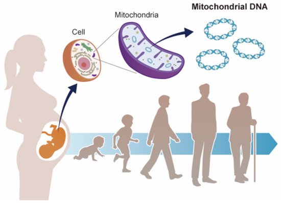
Figure 1), focusing on recent epidemiological studies that suggest a correlation between environmental factors and mtDNA copy number changes during the fetal period.
Figure 1. Mitochondrial DNA and developmental origins of health and disease (DOHaD). Mitochondrial DNA copy number increases or decreases in response to environmental changes. Environmental stress during fetal life may contribute to the risk of disease in adulthood via mitochondrial DNA copy number.

3. DOHaD Theory

The sum of chemical, physical, and biological stresses from the environment, namely the exposome, contributes to human health conditions [62]. Studies by Barker on infants with low birth weights in the 1980s [63][64][65] led to the development of the Fetal Origins of Adult Disease theory in 1990 [54]. Taking into account the results of subsequent epidemiological studies [66][67], the DOHaD theory was proposed in 2004 [55].
Today, a number of epidemiological studies and animal model studies suggest that environmental factors in embryonic, fetal, neonatal, and early childhood periods are associated with health and disease in adulthood and old age, including the risk of developing chronic diseases, supporting the DOHaD theory [68][69][70][71][72][73][74][75][76][77]. For example, in cases of food deprivation during pregnancy, the fetus acquires an energy-saving constitution in anticipation of the environment after birth, in which nutrient intake may be insufficient [78]. If the mother has a glucose-intolerant pregnancy (gestational diabetes mellitus (GDM)) or is obese, it can also cause obesity in the child. It is thought that the fetus receives an excess supply of glucose from the mother, which causes insulin to be overproduced. In addition, the growth-promoting effects of insulin and insulin-like growth factors (IGFs) may be the cause of obese children (gigantism). In addition to nutritional status, various environmental factors, such as exposure to environmental chemicals (e.g., endocrine-disrupting chemicals), are risk factors for disease development [79][80][81]. It is well known that passive smoke exposure during pregnancy interferes with fetal brain development [82]. Low birth weight, especially intrauterine growth retardation, is known to be a risk factor for chronic kidney disease and hypertension. The kidneys are made up of nephrons (renal units), and there are usually around 2 million nephrons per kidney. However, low maternal nutrition, infections, and drugs (ethanol, gentamicin, NSAIDs) can cause the number of nephrons in the fetus to be extremely low, which may lead to the development of hypertension and chronic kidney disease after birth [83]. Other factors such as delayed growth outside the uterus and postnatal administration of steroids and antimicrobials have also been suggested to be associated with the risk of decreased renal function, leading to renal disease. According to research, circadian rhythms during pregnancy also play a role in the health and well-being of children in adulthood, including their risk of particular diseases [84][85]. In addition, gestational hypertension and gestational diabetes may lead to worse health outcomes in newborns [86][87]. Previous studies have provided support for the roles of both the epigenome and gut microbiota during the early postnatal period in future health, thereby supporting the DOHaD theory [88][89][90][91].
Based on the DOHaD theory, there is believed to be a link between exposure to external stress in the fetal environment and risk of noncommunicable diseases (NCDs) in adulthood [92]. Given the increase in the proportion of the aging population in many developed countries, reducing health expenditure through the prevention of NCDs has become an important social and political issue. Previous research suggested that research focusing on the DOHaD was important to develop regional strategies to contribute to a reduction in the incidence of NCDs in low-income countries [93].

References

  1. Luft, R.; Ikkos, D.; Palmieri, G.; Ernster, L.; Afzerius, B. A case of severe hypermetabolism of nonthyroid origin with a defect in the maintenance of mitochondrial respiratory control: A correlated clinical, biochemical, and morphological study. J. Clin. Investig. 1962, 41, 1776–1804.
  2. Roger, A.J.; Muñoz-Gómez, S.A.; Kamikawa, R. The Origin and Diversification of Mitochondria. Curr. Biol. 2017, 27, R1177–R1192.
  3. Brillo, V.; Chieregato, L.; Leanza, L.; Muccioli, S.; Costa, R. Mitochondrial Dynamics, ROS, and Cell Signaling: A Blended Overview. Life 2021, 11, 332.
  4. Ng, Y.S.; Turnbull, D.M. Mitochondrial disease: Genetics and management. J. Neurol. 2016, 263, 179–191.
  5. Gibson, K.; Halliday, J.L.; Kirby, D.M.; Yaplito-Lee, J.; Thorburn, D.R.; Boneh, A. Mitochondrial oxidative phosphorylation disorders presenting in neonates: Clinical manifestations and enzymatic and molecular diagnoses. Pediatrics 2008, 122, 1003–1008.
  6. Ohtake, A.; Murayama, K.; Mori, M.; Harashima, H.; Yamazaki, T.; Tamaru, S.; Yamashita, Y.; Kishita, Y.; Nakachi, Y.; Kohda, M.; et al. Diagnosis and molecular basis of mitochondrial respiratory chain disorders: Exome sequencing for disease gene identification. Biochim. Biophys. Acta 2014, 1840, 1355–1359.
  7. Kohda, M.; Tokuzawa, Y.; Kishita, Y.; Nyuzuki, H.; Moriyama, Y.; Mizuno, Y.; Hirata, T.; Yatsuka, Y.; Yamashita-Sugahara, Y.; Nakachi, Y.; et al. A Comprehensive Genomic Analysis Reveals the Genetic Landscape of Mitochondrial Respiratory Chain Complex Deficiencies. PLoS Genet. 2016, 12, e1005679.
  8. Pavlakis, S.G.; Phillips, P.C.; DiMauro, S.; de Vivo, D.C.; Rowland, L.P. Mitochondrial myopathy, encephalopathy, lactic acidosis, and strokelike episodes: A distinctive clinical syndrome. Ann. Neurol. 1984, 16, 481–488.
  9. Goto, Y.I.; Nonaka, I.; Horai, S. A mutation in the tRNALeu(UUR) gene associated with the MELAS subgroup of mitochondrial encephalomyopathies. Nature 1990, 348, 651–653.
  10. Yatsuga, S.; Povalko, N.; Nishioka, J.; Katayama, K.; Kakimoto, N.; Matsuishi, T.; Kakuma, T.; Koga, Y. MELAS: A nationwide prospective cohort study of 96 patients in Japan. Biochim. Biophys. Acta Gen. Subj. 2012, 1820, 619–624.
  11. Fukuhara, N.; Tokiguchi, S.; Shirakawa, K.; Tsubaki, T. Myoclonus epilepsy associated with ragged-red fibres (mitochondrial abnormalities): Disease entity or a syndrome? Light- and electron-microscopic studies of two cases and review of literature. J. Neurol. Sci. 1980, 47, 117–133.
  12. Shoffner, J.M.; Lott, M.T.; Lezza, A.M.S.; Seibel, P.; Ballinger, S.W.; Wallace, D.C. Myoclonic epilepsy and ragged-red fiber disease (MERRF) is associated with a mitochondrial DNA tRNALys mutation. Cell 1990, 61, 931–937.
  13. Holt, I.J.; Harding, A.E.; Cooper, J.M.; Schapira, A.H.V.; Toscano, A.; Clark, J.B.; Morgan-Hughes, J.A. Mitochondrial myopathies: Clinical and biochemical features of 30 patients with major deletions of muscle mitochondrial DNA. Ann. Neurol. 1989, 26, 699–708.
  14. Moraes, C.T.; DiMauro, S.; Zeviani, M.; Lombes, A.; Shanske, S.; Miranda, A.F.; Nakase, H.; Bonilla, E.; Werneck, L.C.; Servidei, S.; et al. Mitochondrial DNA Deletions in Progressive External Ophthalmoplegia and Kearns-Sayre Syndrome. N. Engl. J. Med. 1989, 320, 1293–1299.
  15. Khambatta, S.; Nguyen, D.L.; Beckman, T.J.; Wittich, C.M. Kearns-Sayre syndrome: A case series of 35 adults and children. Int. J. Gen. Med. 2014, 7, 325–332.
  16. López-Otín, C.; Blasco, M.A.; Partridge, L.; Serrano, M.; Kroemer, G. The hallmarks of aging. Cell 2013, 153, 1194–1217.
  17. John, J.C.S. Genomic Balance: Two Genomes Establishing Synchrony to Modulate Cellular Fate and Function. Cells 2019, 8, 1306.
  18. Al Rawi, S.; Louvet-Vallée, S.; Djeddi, A.; Sachse, M.; Culetto, E.; Hajjar, C.; Boyd, L.; Legouis, R.; Galy, V. Postfertilization autophagy of sperm organelles prevents paternal mitochondrial DNA transmission. Science 2011, 334, 1144–1147.
  19. Sato, M.; Sato, K. Degradation of paternal mitochondria by fertilization-triggered autophagy in C. elegans embryos. Science 2011, 334, 1141–1144.
  20. Killackey, S.A.; Philpott, D.J.; Girardin, S.E. Mitophagy pathways in health and disease. J. Cell Biol. 2020, 219, e202004029.
  21. Onishi, M.; Yamano, K.; Sato, M.; Matsuda, N.; Okamoto, K. Molecular mechanisms and physiological functions of mitophagy. EMBO J. 2021, 40, e104705.
  22. Rossignol, R.; Faustin, B.; Rocher, C.; Malgat, M.; Mazat, J.P.; Letellier, T. Mitochondrial threshold effects. Biochem. J. 2003, 370, 751–762.
  23. Nissanka, N.; Moraes, C.T. Mitochondrial DNA heteroplasmy in disease and targeted nuclease-based therapeutic approaches. EMBO Rep. 2020, 21, e49612.
  24. Pohjoismäki, J.L.O.; Goffart, S.; Tyynismaa, H.; Willcox, S.; Ide, T.; Kang, D.; Suomalainen, A.; Karhunen, P.J.; Griffith, J.D.; Holt, I.J.; et al. Human heart mitochondrial DNA is organized in complex catenated networks containing abundant four-way junctions and replication forks. J. Biol. Chem. 2009, 284, 21446–21457.
  25. Mercer, T.R.; Neph, S.; Dinger, M.E.; Crawford, J.; Smith, M.A.; Shearwood, A.M.J.; Haugen, E.; Bracken, C.P.; Rackham, O.; Stamatoyannopoulos, J.A.; et al. The human mitochondrial transcriptome. Cell 2011, 146, 645–658.
  26. Barchiesi, A.; Vascotto, C. Transcription, processing, and decay of mitochondrial RNA in health and disease. Int. J. Mol. Sci. 2019, 20, 2221.
  27. Wei, W.; Tuna, S.; Keogh, M.J.; Smith, K.R.; Aitman, T.J.; Beales, P.L.; Bennett, D.L.; Gale, D.P.; Bitner-Glindzicz, M.A.K.; Black, G.C.; et al. Germline selection shapes human mitochondrial DNA diversity. Science 2019, 364, eaau6520.
  28. Schaefer, A.M.; Taylor, R.W.; Turnbull, D.M.; Chinnery, P.F. The epidemiology of mitochondrial disorders—Past, present and future. Biochim. Biophys. Acta 2004, 1659, 115–120.
  29. Schaefer, A.M.; McFarland, R.; Blakely, E.L.; He, L.; Whittaker, R.G.; Taylor, R.W.; Chinnery, P.F.; Turnbull, D.M. Prevalence of mitochondrial DNA disease in adults. Ann. Neurol. 2008, 63, 35–39.
  30. Gorman, G.S.; Schaefer, A.M.; Ng, Y.; Gomez, N.; Blakely, E.L.; Alston, C.L.; Feeney, C.; Horvath, R.; Yu-Wai-Man, P.; Chinnery, P.F.; et al. Prevalence of nuclear and mitochondrial DNA mutations related to adult mitochondrial disease. Ann. Neurol. 2015, 77, 753–759.
  31. Murayama, K.; Shimura, M.; Liu, Z.; Okazaki, Y.; Ohtake, A. Recent topics: The diagnosis, molecular genesis, and treatment of mitochondrial diseases. J. Hum. Genet. 2019, 64, 113–125.
  32. Frazier, A.E.; Thorburn, D.R.; Compton, A.G. Mitochondrial energy generation disorders: Genes, mechanisms, and clues to pathology. J. Biol. Chem. 2019, 294, 5386–5395.
  33. Sato, A.; Kono, T.; Nakada, K.; Ishikawa, K.; Inoue, S.I.; Yonekawa, H.; Hayashi, J.I. Gene therapy for progeny of mito-mice carrying pathogenic mtDNA by nuclear transplantation. Proc. Natl. Acad. Sci. USA 2005, 102, 16765–16770.
  34. Yamada, Y.; Harashima, H. Mitochondrial drug delivery systems for macromolecule and their therapeutic application to mitochondrial diseases. Adv. Drug Deliv. Rev. 2008, 60, 1439–1462.
  35. Rai, P.K.; Craven, L.; Hoogewijs, K.; Russell, O.M.; Lightowlers, R.N. Advances in methods for reducing mitochondrial DNA disease by replacing or manipulating the mitochondrial genome. Essays Biochem. 2018, 62, 455–465.
  36. Yamada, Y.; Ito, M.; Arai, M.; Hibino, M.; Tsujioka, T.; Harashima, H. Challenges in promoting mitochondrial transplantation therapy. Int. J. Mol. Sci. 2020, 21, 6365.
  37. Park, A.; Oh, M.; Lee, S.J.; Oh, K.-J.; Lee, E.-W.; Lee, S.C.; Bae, K.-H.; Han, B.S.; Kim, W.K. Mitochondrial Transplantation as a Novel Therapeutic Strategy for Mitochondrial Diseases. Int. J. Mol. Sci. 2021, 22, 4793.
  38. Mengel-From, J.; Thinggaard, M.; Dalgård, C.; Kyvik, K.O.; Christensen, K.; Christiansen, L. Mitochondrial DNA copy number in peripheral blood cells declines with age and is associated with general health among elderly. Hum. Genet. 2014, 133, 1149–1159.
  39. Ashar, F.N.; Moes, A.; Moore, A.Z.; Grove, M.L.; Chaves, P.H.M.; Coresh, J.; Newman, A.B.; Matteini, A.M.; Bandeen-Roche, K.; Boerwinkle, E.; et al. Association of mitochondrial DNA levels with frailty and all-cause mortality. J. Mol. Med. 2015, 93, 177–186.
  40. Dolcini, J.; Kioumourtzoglou, M.-A.; Cayir, A.; Sanchez-Guerra, M.; Brennan, K.J.; Dereix, A.E.; Coull, B.A.; Spiro, A.; Vokonas, P.; Schwartz, J.; et al. Age and mitochondrial DNA copy number influence the association between outdoor temperature and cognitive function. Environ. Epidemiol. 2020, 4, e0108.
  41. Castellani, C.A.; Longchamps, R.J.; Sumpter, J.A.; Newcomb, C.E.; Lane, J.A.; Grove, M.L.; Bressler, J.; Brody, J.A.; Floyd, J.S.; Bartz, T.M.; et al. Mitochondrial DNA copy number can influence mortality and cardiovascular disease via methylation of nuclear DNA CpGs. Genome Med. 2020, 12, 84.
  42. Fazzini, F.; Lamina, C.; Fendt, L.; Schultheiss, U.T.; Kotsis, F.; Hicks, A.A.; Meiselbach, H.; Weissensteiner, H.; Forer, L.; Krane, V.; et al. Mitochondrial DNA copy number is associated with mortality and infections in a large cohort of patients with chronic kidney disease. Kidney Int. 2019, 96, 480–488.
  43. Spinazzola, A.; Invernizzi, F.; Carrara, F.; Lamantea, E.; Donati, A.; DiRocco, M.; Giordano, I.; Meznaric-Petrusa, M.; Baruffini, E.; Ferrero, I.; et al. Clinical and molecular features of mitochondrial DNA depletion syndromes. J. Inherit. Metab. Dis. 2009, 32, 143–158.
  44. Copeland, W.C. Defects in mitochondrial DNA replication and human disease. Crit. Rev. Biochem. Mol. Biol. 2012, 47, 64–74.
  45. Young, M.J.; Copeland, W.C. Human mitochondrial DNA replication machinery and disease. Curr. Opin. Genet. Dev. 2016, 38, 52–62.
  46. Suomalainen, A.; Isohanni, P. Mitochondrial DNA depletion syndromes—Many genes, common mechanisms. Neuromuscul. Disord. 2010, 20, 429–437.
  47. Sun, X.; John, J.C.S. The role of the mtDNA set point in differentiation, development and tumorigenesis. Biochem. J. 2016, 473, 2955–2971.
  48. Gemma, C.; Sookoian, S.; Alvariñas, J.; García, S.I.; Quintana, L.; Kanevsky, D.; González, C.D.; Pirola, C.J. Mitochondrial DNA depletion in small- and large-for-gestational-age newborns. Obesity 2006, 14, 2193–2199.
  49. Barker, D.J.P.; Godfrey, K.M.; Fall, C.; Osmond, C.; Winter, P.D.; Shaheen, S.O. Relation of birth weight and childhood respiratory infection to adult lung function and death from chronic obstructive airways disease. BMJ 1991, 303, 671–675.
  50. Ramadhani, M.K.; Grobbee, D.E.; Bots, M.L.; Cabezas, M.C.; Vos, L.E.; Oren, A.; Uiterwaal, C.S.P.M. Lower birth weight predicts metabolic syndrome in young adults: The Atherosclerosis Risk in Young Adults (ARYA)-study. Atherosclerosis 2006, 184, 21–27.
  51. Geelhoed, J.J.M.; Jaddoe, V.W.V. Early influences on cardiovascular and renal development. Eur. J. Epidemiol. 2010, 25, 677–692.
  52. Memon, A.A.; Sundquist, J.; Hedelius, A.; Palmér, K.; Wang, X.; Sundquist, K. Association of mitochondrial DNA copy number with prevalent and incident type 2 diabetes in women: A population-based follow-up study. Sci. Rep. 2021, 11, 4608.
  53. Yang, S.Y.; Castellani, C.A.; Longchamps, R.J.; Pillalamarri, V.K.; O’Rourke, B.; Guallar, E.; Arking, D.E. Blood-derived mitochondrial DNA copy number is associated with gene expression across multiple tissues and is predictive for incident neurodegenerative disease. Genome Res. 2021, 31, 349–358.
  54. Barker, D.J.P. The fetal and infant origins of adult disease. BMJ 1990, 301, 1111.
  55. Gluckman, P.D.; Hanson, M.A. Living with the past: Evolution, development, and patterns of disease. Science 2004, 305, 1733–1736.
  56. Baird, J.; Jacob, C.; Barker, M.; Fall, C.; Hanson, M.; Harvey, N.; Inskip, H.; Kumaran, K.; Cooper, C. Developmental Origins of Health and Disease: A Lifecourse Approach to the Prevention of Non-Communicable Diseases. Healthcare 2017, 5, 14.
  57. Frankenhuis, W.E.; Nettle, D.; McNamara, J.M. Echoes of Early Life: Recent Insights From Mathematical Modeling. Child Dev. 2018, 89, 1504–1518.
  58. Goyal, D.; Limesand, S.W.; Goyal, R. Epigenetic responses and the developmental origins of health and disease. J. Endocrinol. 2019, 242, T105–T119.
  59. Penkler, M.; Hanson, M.; Biesma, R.; Müller, R. DOHaD in science and society: Emergent opportunities and novel responsibilities. J. Dev. Orig. Health Dis. 2019, 10, 268–273.
  60. Fall, C.H.D.; Kumaran, K. Metabolic programming in early life in humans. Philos. Trans. R. Soc. B Biol. Sci. 2019, 374, 20180123.
  61. Briana, D.D.; Malamitsi-Puchner, A. Perinatal biomarkers implying “Developmental Origins of Health and Disease” consequences in intrauterine growth restriction. Acta Paediatr. Int. J. Paediatr. 2020, 109, 1317–1322.
  62. Vermeulen, R.; Schymanski, E.L.; Barabási, A.L.; Miller, G.W. The exposome and health: Where chemistry meets biology. Science 2020, 367, 392–396.
  63. Barker, D.J.P.; Osmond, C. Infant mortality, childhood nutrition, and ischaemic heart disease in England and Wales. Lancet 1986, 327, 1077–1081.
  64. Barker, D.J.P.; Osmond, C.; Winter, P.D.; Margetts, B.; Simmonds, S.J. Weight in infancy and death from ischaemic heart disease. Lancet 1989, 334, 577–580.
  65. Barker, D.J.P.; Bull, A.R.; Osmond, C.; Simmonds, S.J. Fetal and placental size and risk of hypertension in adult life. BMJ 1990, 301, 259–262.
  66. Barker, D.J.P.; Hales, C.N.; Fall, C.H.D.; Osmond, C.; Phipps, K.; Clark, P.M.S. Type 2 (non-insulin-dependent) diabetes mellitus, hypertension and hyperlipidaemia (syndrome X): Relation to reduced fetal growth. Diabetologia 1993, 36, 62–67.
  67. Hofman, P.L.; Regan, F.; Jackson, W.E.; Jefferies, C.; Knight, D.B.; Robinson, E.M.; Cutfield, W.S. Premature Birth and Later Insulin Resistance. N. Engl. J. Med. 2004, 351, 2179–2186.
  68. Barker, D.J.P. The origins of the developmental origins theory. J. Intern. Med. 2007, 261, 412–417.
  69. Roseboom, T.; de Rooij, S.; Painter, R. The Dutch famine and its long-term consequences for adult health. Early Hum. Dev. 2006, 82, 485–491.
  70. Eriksson, J.G. Developmental Origins of Health and Disease—From a small body size at birth to epigenetics. Ann. Med. 2016, 48, 456–467.
  71. Hanson, M. The birth and future health of DOHaD. J. Dev. Orig. Health Dis. 2015, 6, 434–437.
  72. Hsu, C.N.; Lin, Y.J.; Yu, H.R.; Lin, I.C.; Sheen, J.M.; Huang, L.T.; Tain, Y.L. Protection of male rat offspring against hypertension programmed by prenatal dexamethasone administration and postnatal high-fat diet with the Nrf2 activator dimethyl fumarate during pregnancy. Int. J. Mol. Sci. 2019, 20, 3957.
  73. Hsu, C.N.; Lin, Y.J.; Tain, Y.L. Maternal exposure to bisphenol a combined with high-fat diet-induced programmed hypertension in adult male rat offspring: Effects of resveratrol. Int. J. Mol. Sci. 2019, 20, 4382.
  74. Larsen, T.D.; Sabey, K.H.; Knutson, A.J.; Gandy, T.C.T.; Louwagie, E.J.; Lauterboeck, L.; Mdaki, K.S.; Baack, M.L. Diabetic pregnancy and maternal high-fat diet impair mitochondrial dynamism in the developing fetal rat heart by sex-specific mechanisms. Int. J. Mol. Sci. 2019, 20, 3090.
  75. Hofstee, P.; Cuffe, J.S.M.; Perkins, A.V. Analysis of selenoprotein expression in response to dietary selenium deficiency during pregnancy indicates tissue specific differential expression in mothers and sex specific changes in the fetus and offspring. Int. J. Mol. Sci. 2020, 21, 2210.
  76. Alexandre-Gouabau, M.C.; David-Sochard, A.; Royer, A.L.; Parnet, P.; Paillé, V. Moderate high caloric maternal diet impacts dam breast milk metabotype and offspring lipidome in a sex-specific manner. Int. J. Mol. Sci. 2020, 21, 5428.
  77. Visker, J.R.; Dangott, L.J.; Leszczynski, E.C.; Ferguson, D.P. Postnatal growth restriction in mice alters cardiac protein composition and leads to functional impairment in adulthood. Int. J. Mol. Sci. 2020, 21, 9459.
  78. Hales, C.N.; Barker, D.J.P. The thrifty phenotype hypothesis. Br. Med. Bull. 2001, 60, 5–20.
  79. Heindel, J.J.; Skalla, L.A.; Joubert, B.R.; Dilworth, C.H.; Gray, K.A. Review of developmental origins of health and disease publications in environmental epidemiology. Reprod. Toxicol. 2017, 68, 34–48.
  80. Almeida, D.L.; Pavanello, A.; Saavedra, L.P.; Pereira, T.S.; de Castro-Prado, M.A.A.; de Freitas Mathias, P.C. Environmental monitoring and the developmental origins of health and disease. J. Dev. Orig. Health Dis. 2019, 10, 608–615.
  81. Street, M.E.; Bernasconi, S. Endocrine-disrupting chemicals in human fetal growth. Int. J. Mol. Sci. 2020, 21, 1430.
  82. Chen, R.; Clifford, A.; Lang, L.; Anstey, K.J. Is exposure to secondhand smoke associated with cognitive parameters of children and adolescents?—A systematic literature review. Ann. Epidemiol. 2013, 23, 652–661.
  83. Brenner, B.M.; Chertow, G.M. Congenital Oligonephropathy and the Etiology of Adult Hypertension and Progressive Renal Injury. Am. J. Kidney Dis. 1994, 23, 171–175.
  84. Tain, Y.L.; Huang, L.T.; Hsu, C.N. Developmental programming of adult disease: Reprogramming by melatonin? Int. J. Mol. Sci. 2017, 18, 426.
  85. Hsu, C.N.; Tain, Y.L. Light and circadian signaling pathway in pregnancy: Programming of adult health and disease. Int. J. Mol. Sci. 2020, 21, 2232.
  86. Iwama, N.; Metoki, H.; Ohkubo, T.; Ishikuro, M.; Obara, T.; Kikuya, M.; Yagihashi, K.; Nishigori, H.; Sugiyama, T.; Sugawara, J.; et al. Maternal clinic and home blood pressure measurements during pregnancy and infant birth weight: The BOSHI study. Hypertens. Res. 2016, 39, 151–157.
  87. Aisa, M.C.; Cappuccini, B.; Barbati, A.; Clerici, G.; Torlone, E.; Gerli, S.; Di Renzo, G.C. Renal Consequences of Gestational Diabetes Mellitus in Term Neonates: A Multidisciplinary Approach to the DOHaD Perspective in the Prevention and Early Recognition of Neonates of GDM Mothers at Risk of Hypertension and Chronic Renal Diseases in Later Life. J. Clin. Med. 2019, 8, 429.
  88. Gluckman, P.D.; Hanson, M.A.; Buklijas, T.; Low, F.M.; Beedle, A.S. Epigenetic mechanisms that underpin metabolic and cardiovascular diseases. Nat. Rev. Endocrinol. 2009, 5, 401–408.
  89. Chmurzynska, A. Fetal programming: Link between early nutrition, DNA methylation, and complex diseases. Nutr. Rev. 2010, 68, 87–98.
  90. Safi-Stibler, S.; Gabory, A. Epigenetics and the Developmental Origins of Health and Disease: Parental environment signalling to the epigenome, critical time windows and sculpting the adult phenotype. Semin. Cell Dev. Biol. 2020, 97, 172–180.
  91. Sarkar, A.; Yoo, J.Y.; Valeria Ozorio Dutra, S.; Morgan, K.H.; Groer, M. The Association between Early-Life Gut Microbiota and Long-Term Health and Diseases. J. Clin. Med. 2021, 10, 459.
  92. Rodriguez-Caro, H.; Williams, S.A. Strategies to reduce non-communicable diseases in the offspring: Negative and positive in utero programming. J. Dev. Orig. Health Dis. 2018, 9, 642–652.
  93. Tu’akoi, S.; Vickers, M.H.; Bay, J.L. DOHaD in low- and middle-income countries: A systematic review exploring gaps in DOHaD population studies. J. Dev. Orig. Health Dis. 2020, 11, 557–563.
More
Information
Contributor MDPI registered users' name will be linked to their SciProfiles pages. To register with us, please refer to https://encyclopedia.pub/register :
View Times: 644
Revisions: 2 times (View History)
Update Date: 19 Jul 2021
1000/1000