Submitted Successfully!
To reward your contribution, here is a gift for you: A free trial for our video production service.
Thank you for your contribution! You can also upload a video entry or images related to this topic.
Version Summary Created by Modification Content Size Created at Operation
1 + 2149 word(s) 2149 2021-06-30 09:44:27 |
2 format correct -21 word(s) 2128 2021-07-02 02:51:48 |

Video Upload Options

Do you have a full video?

Confirm

Are you sure to Delete?
Cite
If you have any further questions, please contact Encyclopedia Editorial Office.
Umakanthan, S. Genetics in Epstein–Barr Virus-Associated Malignancies. Encyclopedia. Available online: https://encyclopedia.pub/entry/11572 (accessed on 23 April 2024).
Umakanthan S. Genetics in Epstein–Barr Virus-Associated Malignancies. Encyclopedia. Available at: https://encyclopedia.pub/entry/11572. Accessed April 23, 2024.
Umakanthan, Srikanth. "Genetics in Epstein–Barr Virus-Associated Malignancies" Encyclopedia, https://encyclopedia.pub/entry/11572 (accessed April 23, 2024).
Umakanthan, S. (2021, July 01). Genetics in Epstein–Barr Virus-Associated Malignancies. In Encyclopedia. https://encyclopedia.pub/entry/11572
Umakanthan, Srikanth. "Genetics in Epstein–Barr Virus-Associated Malignancies." Encyclopedia. Web. 01 July, 2021.
Genetics in Epstein–Barr Virus-Associated Malignancies
Edit

Global genomic studies have detected the role of genomic alterations in the pathogenesis of Epstein–Barr virus (EBV)-associated tumors. EBV oncoproteins cause a vital shift of EBV from an infectious virus to an oncogenic form during the latent and lytic phase within the lymphoid B cells and epithelial cells. This epigenetic alteration modulates the virus and host genomes and inactivates and disrupts numerous tumor suppressors and signaling pathways. Genomic profiling has played the main role in identifying EBV cancer pathogenesis and its related targeted therapies. 

Epstein–Barr virus genome pathogenesis lymphomas carcinomas

1. Introduction

Epstein–Barr virus (EBV) is a lymphotropic virus belonging to the human herpesvirus family. Based on the epidemiological data, EBV infects more than 90% of the world’s population [1]. It was first discovered in the cells of an African patient suffering from Burkitt lymphoma in 1964. The primary infection occurs typically in children and is asymptomatic [2]. As the infection recurs in the adult stage, the infection manifests as infectious mononucleosis. EBV has demonstrated the transformation of resting B cells into lymphoblastoid oncogenic cell lines resulting in the origin of various lymphomas and epithelial malignancies [3].
The genomic variation in EBV promotes the virus transformation into oncogenic cell lines that are responsible for its tumorigenic outcome [4]. Infectious mononucleosis results from the mutagenic variation of HLA-class I, Burkitt lymphoma arises from B cell proliferation due to MYC translocation, Hodgkin’s lymphoma is caused due to the critical role of NF-Kb, EBV produces “trogocytosis” in the development of NK/T cell lymphoma and nasopharyngeal carcinoma occurs due to functional polymorphism of MAP2K4 suppressor gene [5][6][7].

3. Genomic Structure

EBV genome is composed of 172 kbp linear, double-stranded DNA. The open reading frames (ORF) are involved in encoding proteins for DNA replication, gene expression, and conserving genomic integrity. These coding genes have structural and non-structural functions depending on the ORF sizes [8]. The microRNAs (miRNAs), which include BART and BHRF cluster, are expressed during the various phases of EBV infection and are encoded by the EBV. These miRNAs promote the latent phase of the virus and also target the cellular genes causing carcinomas of various types. The 3′ untranslated region (UTR) in the host genes promotes cancer susceptibility and patient prognosis [9].
Genome sequences reported from previous studies are as follows: (1) B95-8, derived from a case of infectious mononucleosis in North America; (2) WT-EBV, is a more complete reference genome constructed by using B95-8 as its backbone; (3) AG876, derived from a case of Burkitt lymphoma in Western Africa is unique for being the only complete established type 2 genomic sequence of EBV; (4) GD1, (5) GD2, and (6) HKNPC1 are derived from nasopharyngeal carcinoma patients; (7) Akata, derived from Burkitt lymphoma case in Japan; (8) Mutu, derived from a Burkitt lymphoma case in Kenya using NGS technology; (9) C666-1; (10) Raji; (11) K4412-Mi; (12) K4123-Mi; (13) EBVaGC1-9 have also shown regional variations [10][11][12][13].

4. Infection Cycle

During the initial stages, EBV spreads through the saliva and affects the tonsils. The naïve B cells and the follicular dendritic cells in the tonsils bind with the surface glycoprotein of the virus through the complement receptor CD21 [14]. This further activates the B cells, and the virus catalyzes the differentiation of the normal cellular pathway of the B cell through the expression of the viral latent proteins: namely, Epstein–Barr nuclear antigens (EBNA) and latent membrane proteins (LMP). Following this differentiation, the B blast enters the germinal center causing down-regulation of the EBNA proteins. This propels the infected B cell into the germinal center, before exiting into the circulating blood as a memory B cell [15]. This latent inactive state is due to the lack of the virus protein expression. Direct infection of either naïve B cells or memory B cells and also marginal zone memory B cells results in the release of terminal differentiation signals triggering a lytic reactivation of the latent virus [16]. This reactivation phase is subdivided into immediate-early, early, and late phases. The infectious virions are now released into saliva for the transmission of the virus to new hosts, thus concluding the infectious cycle [17].

5. EBV Associated Human Diseases

EBV primarily causes infectious mononucleosis (IM) and also associated with the development of systemic disorders like rheumatoid arthritis, multiple sclerosis, chronic fatigue syndrome, and Vitamin D deficiency [18]. Malignancies caused by EBV include Burkitt lymphoma, Hodgkin’s lymphoma, NK/T-cell lymphoma, nasopharyngeal carcinoma, gastric carcinoma, and breast carcinoma [19]. The genetic origin of these individual lesions along with diagnostic and treatment aspects are reviewed. Since IM forms the basis of EBV oncogenic transformation, IM is included in this review along with other malignancies.

6. Infectious Mononucleosis (IM)

IM is predominantly caused by EBV through the saliva of an infected person and is transmitted by coughing and kissing. It commonly affects adolescents and young adults [20]. EBV viral concentration is detected in the saliva at a wide range of concentrations, peaking during the acute phase. EBV infects the oropharyngeal epithelial cells and the resting B-cells, following which the virus replication is initiated. This period is devoted to incubation, lasting from four to eight weeks. The next phase involves activation of cytotoxic T-lymphocytes and the natural killer cells, inducing a cell-mediated immunity [21]. Reactivation can occur during the later period of life elicited by host immunosuppression events such as infections (e.g., HIV), medications, and surgical transplants [22].
EBV-induced host genetic variation has been highlighted by many gene studies. IM- associated genomic regions and polymorphism of HLA-class I have a higher risk of developing IM. Recent studies have further included homozygous allele 1 of STR D6S510 and STR D6S265 in the high-risk list for developing IM [21][23]. Genomic sequencing has identified certain genetic disorders that are susceptible to develop EBV-induced IM. These include CORO1A, XMEN, ITK, and PRKCD deficiency. CORO1A deficiency causes primary immunodeficiency, XMEN deficiency leads to general immunodeficiency, ITK deficiency causes fatal B-cell proliferation, and PRKCD causes autoimmune lymphoproliferative syndrome [21][22][24].
Clinical features include fever, pharyngitis, cervical lymphadenopathy, tonsillitis, and palatal petechiae. Laboratory evaluation shows lymphocytosis and elevated alanine aminotransferase [25]. 85% of EBV infected patients show a positive Monospot test (heterophile antibody test): a famous laboratory test, owing to its rapid diagnosis and low cost. VCA IgM antibody is specific during the early infectious period and VCA IgG antibody is best for past infection diagnosis. Both these tests are not usually performed at the point of care sites [26][27]. Treatment for IM mainly focuses on symptomatic relief by rest, fluid replacement therapy, antipyretics, and analgesics. Antiviral drugs like acyclovir, valacyclovir, and steroid usage have been of limited clinical benefit [28].

7. Burkitt Lymphoma (BL)

BL is classified into endemic, sporadic, and HIV-associated types. EBV is associated with over 90% of endemic BL and is prevalent in regions where malaria is hyper-endemic such as in the equatorial African regions [29]. HIV-associated types are EBV positive in approximately 30%. Endemic EBV-associated BL is common in children and often presents with involvement of the jaw, facial bones, renal, and extranodal sites [30].
The B cells infected by the EBV along with MYC translocation and co-factors such as malaria and HIV infection initiates B cell activation and proliferation. As the number of B cell infections rises, there is germinal center expansion and accumulation of oncogenic mutation [31]. The coupling effect of malaria-mediated activation and germinal center B cell proliferation results in the formation of BL progenitor cells. The vital proliferative and apoptotic pathways include gene activating and gene inhibitory mutations. The gene activating mutations are the c-MYC, TCF3, and CCND3. The gene inhibitory mutations include P53, PTEN, ID3, and CDKN2A [32][33].
Histologically this tumor type exhibits diffuse effacement of the nodal architecture replaced by medium-sized monomorphic tumor cells with round nuclei and numerous nucleoli. Due to their high proliferation rate, presence of numerous atypical mitosis, macrophages, and apoptotic debris, they display a striking “starry sky” pattern (Figure 1A,B). The diagnosis of BL is dependent solely on its genetic and immunophenotype analysis such as CD10, CD 20, and high Mib-1 labeling index (Figure 1C) [31][34].
Figure 1. Burkitt Lymphoma: (A) Diffuse effacement of the nodal architecture by atypical lymphoid cells. (B) Starry sky pattern (C) high Mib-1 labeling index.
However, imaging studies contribute to the evaluation of the clinical course, treatment response, and complications of BL [35]. This aggressive lymphoma is treated with multi-drug chemotherapy and immunotherapy. The ID3/EZA/cyclinD3 pathway targeted therapy and inhibitors of the c-MYC, PI3 kinase and Bcl2 family are under detailed genetic mapping for the development of new therapeutic target agents [36].

8. Hodgkin’s Lymphoma (HL)

Hodgkin’s disease was first described by Sir Thomas Hodgkin in 1832. Since then, there has been controversy on whether to classify it as an inflammatory, infectious, or malignant disease [37]. HL has shown typical bimodal age distribution with EBV+ cases mainly showing an initial peak in children of developing nations. The geographic distribution of EBV+HL cases is across Kenya (92%), China (65%), South and Central America (50–95%), and North America and Europe (20–50%) [38].
EBV infection targets the naïve B cells, interferes with B cell genetic differentiation, and rescues B-cell receptor-deficient germinal center B cells from apoptosis. NF-kB plays a critical role during the rescue process by activating the expression of anti-apoptotic DISC-inhibitor c-FLIP [39]. These EBV-infected HL cells express various LMPs that activate NF-kB. Notch ligation has proven to play a vital role in LMP-1 regulation by inhibiting LMP-1 expression and promoting EBV nuclear antigen -2 (EBNA2) during the primary infectious phase [38][40]. Activated Notch ligation also downregulates LMP2A during the lymphoblastoid transformation of B cells. LMP-1 promotes the expression of the collagen receptor, discoidin domain receptor 1 (DDR 1) that forms a major chronic inflammatory component for the HL microenvironment. LMP-1 expression explains the varied presentation of EBV-infected patients from an asymptomatic phase to a severe potent oncogenic phase. In some HL cases, EBV causes inactivation of A20 tumor suppressor gene alleles [39][41][42].
HIV- infected patients almost always express type II latency of EBV infection (Figure 2A,B). This is characterized by morphological differences by increased CD 163+ spindle-shaped macrophages forming a “sarcomatoid” pattern. This provides a favorable treatment using the cART regime (doxorubicin, bleomycin, vinblastine, dacarbazine) providing an outcome similar to HIV-negative patients [43][44].
Figure 2. Hodgkin’s lymphoma (A) in HIV (B) positive immunostain for Epstein–Barr virus-latent membrane protein.

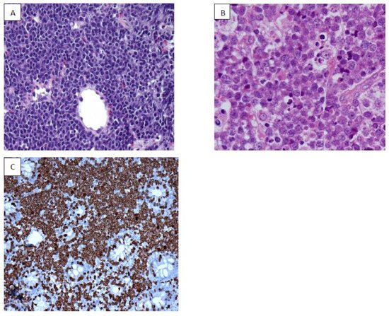
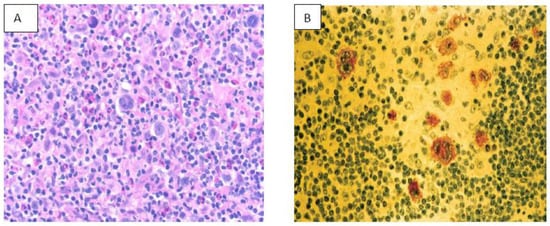
9. NK/T Cell Lymphoma (NKTCL)

EBV is well documented as an oncogenic virus in B cell neoplasm; however, its role in the pathogenesis of NKTCL has been complex. Mature NKTCL is a group of heterogeneous tumors that have complex treatment options, aggressive clinical presentation, and poor prognosis [45]. The geographic distribution of EBV-induced NKTCL has been more concentrated in Asia (China, Korea), South America (Peru, Argentina), Central America (Mexico), and Central Africa. EBV in NKTCL is classified into nodal and extra-nodal types. The role of EBV in angioimmunoblastic T-cell lymphoma, nasal type of extranodal NKTL, and aggressive NK- cell leukemia is well researched and documented [46].
The role of EBV infection in NKTCL has been hypothesized during the targeted killing of EBV-infected cells by NK/T cells. The patients with overlapping evidence of EBV- associated diseases are shown to have circulating EBV-infected NK/T cells (EBV positive NK/T cells) [46][47]. This positivity is due to the effect of the “trogocytosis” phenomenon caused by glycoproteins (gp 350, gp 42, and gp85) and cellular protein (CD 21). This further induces the expression of EBNA (types 1,2,3A,3B,3C) and LMP (1,2A,2B) proteins that result in genetic polymorphisims. LMP1 acts as an active member of tumor necrosis factor (TNF) and downregulates NF-Kb and MAPK signal pathways [45][46][48]. This further causes variation in the C-terminus of LMP1, deletion of 30bp, 33bp repeats, insertion of 15bp, and other amino acid substitutions. These genetic expressions, deletions, and variations have been studied in numerous molecular genetic pathways using whole-genome sequencing, targeted and exome sequencing. This has allowed the discovery of more target-specific therapies to these sets of complex malignancies [46][49]. The most common genetic alteration in EBV-induced NKTCL is the loss of 6q21 (20–43% cases) followed by recurrent losses in chromosomes 1p4 and 5p13. JAK-STAT signaling pathways along with STAT3, KMTZD, and TP53 are the most recurrently mutated genes in EBV-induced NKTCL [50].
Extranodal NKTCL occurs predominantly in midline facial structures and can also affect paranasal sinuses, orbit, jaw, and salivary glands. Symptoms vary from systemic features (fever, weight loss, night sweats) to hemophagocytic disorders [51]. Imaging studies (CT, MRI, PET/CT) coupled with EBV-DNA biomarker assessment are essential for diagnosis [52].
Treatment modality varies on the stage of NKTCL. Stage I/II are treated mainly with radiotherapy. Previous chemotherapeutic regimes using anthracycline-based CHOP (cyclophosphamide, doxorubicin, vincristine, and prednisone) have shown to cause more toxicity and drug resistance [53]. The recently introduced LVP (L-asparaginase, vincristine, and prednisone) and GELOX (gemcitabine, oxaliplatin, and L-asparaginase) are shown to be more effective in this stage [51][54]. Advanced/relapsed/recurrent NKTCL are treated with selective chemotherapy but due to poor host response and drug-induced toxicity has resulted in poor patient prognosis in many of the cases. Hematopoietic stem cell transplant and immunotherapy are still under trial [55].

References

  1. Hjalgrim, H.; Friborg, J.; Melbye, M. The Epidemiology of EBV and its Association with Malignant Disease. In Human Herpesviruses: Biology, Therapy, and Immunoprophylaxis; Arvin, A., Campadelli-Fiume, G., Mocarski, E., Moore, S.P., Roizman, B., Whitley, R., Yamanishi, K., Eds.; Cambridge University Press: Cambridge, UK, 2007; Chapter 53. Available online: (accessed on 22 May 2021).
  2. Abdel-Hamid, M.; Chen, J.-J.; Constantine, N.; Massoud, M.; Raab-Traub, N. EBV strain variation: Geographical distribution and relation to disease state. Virology 1992, 190, 168–175.
  3. IARC Working Group on the Evaluation of Carcinogenic Risks to Humans. Biological Agents. In (IARC Monographs on the Evaluation of Carcinogenic Risks to Humans, No. 100B.) EPSTEIN-BARR VIRUS; International Agency for Research on Cancer: Lyon, France, 2012. Available online: (accessed on 22 May 2021).
  4. Raab-Traub, N. EBV-induced oncogenesis. In Human Herpesviruses: Biology, Therapy, and Immunoprophylaxis; Arvin, A., Campadelli-Fiume, G., Mocarski, E., Moore, S.P., Roizman, B., Whitley, R., Yamanishi, K., Eds.; Cambridge University Press: Cambridge, UK, 2007; Chapter 55. Available online: (accessed on 22 May 2021).
  5. Keating, S.; Prince, S.; Jones, M.; Rowe, M. The Lytic Cycle of Epstein-Barr Virus Is Associated with Decreased Expression of Cell Surface Major Histocompatibility Complex Class I and Class II Molecules. J. Virol. 2002, 76, 8179–8188.
  6. Shannon-Lowe, C.; Rickinson, A.B.; Bell, A.I. Epstein–Barr virus-associated lymphomas. Philos. Trans. R. Soc. B Biol. Sci. 2017, 372, 20160271.
  7. Münz, M. Dendritic cells during Epstein Barr virus infection. Front. Microbiol. 2014, 5, 308.
  8. Young, L.S.; Arrand, J.R.; Murray, P.G. EBV gene expression and regulation. In Human Herpesviruses: Biology, Therapy, and Immunoprophylaxis; Arvin, A., Campadelli-Fiume, G., Mocarski, E., Moore, S.P., Roizman, B., Whitley, R., Yamanishi, K., Eds.; Cambridge University Press: Cambridge, UK, 2007; Chapter 27. Available online: (accessed on 22 May 2021).
  9. Sunagawa, K.; Hishima, T.; Fukumoto, H.; Hasegawa, H.; Katano, H. Conserved sequences of BART and BHRF regions encoding viral microRNAs in Epstein-Barr virus-associated lymphoma. BMC Res. Notes 2017, 10, 279.
  10. Weiss, E.R.; Lamers, S.L.; Henderson, J.L.; Melnikov, A.; Somasundaran, M.; Garber, M.; Selin, L.; Nusbaum, C.; Luzuriaga, K. Early Epstein-Barr Virus Genomic Diversity and Convergence toward the B95.8 Genome in Primary Infection. J. Virol. 2018, 92, e01466-17.
  11. Lei, H.; Li, T.; Hung, G.-C.; Li, B.; Tsai, S.; Lo, S.-C. Identification and characterization of EBV genomes in spontaneously immortalized human peripheral blood B lymphocytes by NGS technology. BMC Genom. 2013, 14, 804.
  12. Kwok, H.; Tong, A.H.Y.; Lin, C.H.; Lok, S.; Farrell, P.; Kwong, D.L.W.; Chiang, A.K.S. Genomic Sequencing and Comparative Analysis of Epstein-Barr Virus Genome Isolated from Primary Nasopharyngeal Carcinoma Biopsy. PLoS ONE 2012, 7, e36939.
  13. Abate, F.; Ambrosio, M.R.; Mundo, L.; Laginestra, M.A.; Fuligni, F.; Rossi, M.; Zairis, S.; Gazaneo, S.; De Falco, G.; Lazzi, S.; et al. Distinct Viral and Mutational Spectrum of Endemic Burkitt Lymphoma. PLoS Pathog. 2015, 11, e1005158.
  14. Wohlford, E.M.; Baresel, P.C.; Wilmore, J.R.; Mortelliti, A.J.; Coleman, C.B.; Rochford, R. Changes in Tonsil B Cell Phenotypes and EBV Receptor Expression in Children under 5-Years-Old. Cytom. Part B Clin. Cytom. 2018, 94, 291–301.
  15. Jog, N.; McClain, M.T.; Heinlen, L.D.; Gross, T.; Towner, R.; Guthridge, J.M.; Axtell, R.C.; Pardo, G.; Harley, J.B.; James, J.A. Epstein Barr virus nuclear antigen 1 (EBNA-1) peptides recognized by adult multiple sclerosis patient sera induce neurologic symptoms in a murine model. J. Autoimmun. 2020, 106, 102332.
  16. Frappier, L. The Epstein-Barr Virus EBNA1 Protein. Scientifica 2012, 2012, 438204.
  17. De França, T.R.T.; Carvalho, A.D.A.T.; Gomes, V.B.; Gueiros, L.A.; Porter, S.R.; Leão, J.C. Salivary shedding of Epstein–Barr virus and cytomegalovirus in people infected or not by human immunodeficiency virus 1. Clin. Oral Investig. 2011, 16, 659–664.
  18. Houen, G.; Trier, N.H. Epstein-Barr Virus and Systemic Autoimmune Diseases. Front. Immunol. 2021, 11, 7380.
  19. Ko, Y.-H. EBV and human cancer. Exp. Mol. Med. 2015, 47, e130.
  20. Balfour, H.H., Jr.; Dunmire, S.K.; Hogquist, K.A. Infectious mononucleosis. Clin. Transl. Immunol. 2015, 4, e33.
  21. Aslan, N.; Watkin, L.B.; Gil, A.; Mishra, R.; Clark, F.G.; Welsh, R.M.; Ghersi, D.; Luzuriaga, K.; Selin, L.K. Severity of Acute Infectious Mononucleosis Correlates with Cross-Reactive Influenza CD8 T-Cell Receptor Repertoires. MBio 2017, 8, e01841-17.
  22. Gequelin, L.C.F.; Riediger, I.N.; Nakatani, S.M.; Biondo, A.W.; Bonfim, C.M. Epstein-Barr virus: General factors, virus-related diseases and measurement of viral load after transplant. Rev Bras Hematol Hemoter. Rev. Bras. Hematol. Hemoter. 2011, 33, 383–388.
  23. Houldcroft, C.J.; Kellam, P. Host genetics of Epstein–Barr virus infection, latency and disease. Rev. Med. Virol. 2015, 25, 71–84.
  24. Li, F.-Y.; Chaigne-Delalande, B.; Kanellopoulou, C.; Davis, J.C.; Matthews, H.F.; Douek, D.C.; Cohen, J.I.; Uzel, G.; Su, H.C.; Lenardo, M.J. Second messenger role for Mg2+ revealed by human T-cell immunodeficiency. Nature 2011, 475, 471–476.
  25. Fugl, A.; Andersen, C.L. Epstein-Barr virus and its association with disease—A review of relevance to general practice. BMC Fam. Pract. 2019, 20, 62.
  26. Marshall-Andon, T.; Heinz, P. How to use … the Monospot and other heterophile antibody tests. Arch. Dis. Child. Educ. Pract. Ed. 2017, 102, 188–193.
  27. Sohn, M.J.; Cho, J.M.; Moon, J.S.; Ko, J.S.; Yang, H.R. EBV VCA IgM and cytomegalovirus IgM dual positivity is a false positive finding related to age and hepatic involvement of primary Epstein–Barr virus infection in children. Medicine 2018, 97, e12380.
  28. Stanfield, B.A.; Luftig, M.A. Recent advances in understanding Epstein-Barr virus. F1000Research 2017, 6, 386.
  29. Hämmerl, L.; Colombet, M.; Rochford, R.; Ogwang, D.M.; Parkin, D.M. The burden of Burkitt lymphoma in Africa. Infect. Agents Cancer 2019, 14, 17.
  30. Kalisz, K.; Alessandrino, F.; Beck, R.; Smith, D.; Kikano, E.; Ramaiya, N.H.; Tirumani, S.H. An update on Burkitt lymphoma: A review of pathogenesis and multimodality imaging assessment of disease presentation, treatment response, and recurrence. Insights Imaging 2019, 10, 56.
  31. Bouska, A.; Bi, C.; Lone, W.; Zhang, W.; Kedwaii, A.; Heavican, T.; Lachel, C.M.; Yu, J.; Ferro, R.; Eldorghamy, N.; et al. Adult high-grade B-cell lymphoma with Burkitt lymphoma signature: Genomic features and potential therapeutic targets. Blood 2017, 130, 1819–1831.
  32. Umakanthan, S.; Bukelo, M.M. Concise genetic profile of lung carcinoma. Postgrad Med. J. 2021.
  33. Yu, L.; Yu, T.-T.; Young, K.H. Cross-talk between Myc and p53 in B-cell lymphomas. Chronic Dis. Transl. Med. 2019, 5, 139–154.
  34. Chuang, S.-S.; Ye, H.; Du, M.-Q.; Lu, C.-L.; Dogan, A.; Hsieh, P.-P.; Huang, W.-T.; Jung, Y.-C. Histopathology and Immunohistochemistry in Distinguishing Burkitt Lymphoma from Diffuse Large B-Cell Lymphoma with Very High Proliferation Index and with or without a Starry-Sky Pattern: A comparative study with EBER and FISH. Am. J. Clin. Pathol. 2007, 128, 558–564.
  35. Oosten, L.E.M.; Chamuleau, M.E.D.; Thielen, F.W.; De Wreede, L.C.; Siemes, C.; Doorduijn, J.K.; Smeekes, O.S.; Kersten, M.J.; Hardi, L.; Baars, J.W.; et al. Treatment of sporadic Burkitt lymphoma in adults, a retrospective comparison of four treatment regimens. Ann. Hematol. 2018, 97, 255–266.
  36. Schmitz, R.; Young, R.M.; Ceribelli, M.; Jhavar, S.; Xiao, W.; Zhang, M.; Wright, G.L.; Shaffer, A.L.; Hodson, D.J.; Buras, E.; et al. Burkitt lymphoma pathogenesis and therapeutic targets from structural and functional genomics. Nat. Cell Biol. 2012, 490, 116–120.
  37. Stone, M.J. Thomas Hodgkin: Medical Immortal and Uncompromising Idealist. In Baylor University Medical Center Proceedings; Informa UK Limited: London, UK, 2005; Volume 18, pp. 368–375.
  38. Salati, M.; Cesaretti, M.; Macchia, M.; El Mistiri, M.; Federico, M. Epidemiological Overview of Hodgkin Lymphoma across the Mediterranean Basin. Mediterr. J. Hematol. Infect. Dis. 2014, 6, e2014048.
  39. Massini, G.; Siemer, D.; Hohaus, S. EBV in Hodgkin Lymphoma. Mediterr. J. Hematol. Infect. Dis. 2009, 1, e2009013.
  40. Arruga, F.; Vaisitti, T.; Deaglio, S. The NOTCH Pathway and Its Mutations in Mature B Cell Malignancies. Front. Oncol. 2018, 8, 550.
  41. Vrzalikova, K.; Sunmonu, T.; Reynolds, G.; Murray, P. Contribution of Epstein–Barr Virus Latent Proteins to the Pathogenesis of Classical Hodgkin Lymphoma. Pathogens 2018, 7, 59.
  42. Tan, G.W.; Visser, L.; Tan, L.P.; Berg, A.V.D.; Diepstra, A. The Microenvironment in Epstein–Barr Virus-Associated Malignancies. Pathogens 2018, 7, 40.
  43. Schmitz, R.; Hansmann, M.-L.; Bohle, V.; Martin-Subero, J.I.; Hartmann, S.; Mechtersheimer, G.; Klapper, W.; Vater, I.; Giefing, M.; Gesk, S.; et al. TNFAIP3 (A20) is a tumor suppressor gene in Hodgkin lymphoma and primary mediastinal B cell lymphoma. J. Exp. Med. 2009, 206, 981–989.
  44. Carbone, A.; Vaccher, E.; Gloghini, A.; Pantanowitz, L.; Abayomi, A.; De Paoli, P.; Franceschi, S. Diagnosis and management of lymphomas and other cancers in HIV-infected patients. Nat. Rev. Clin. Oncol. 2014, 11, 223–238.
  45. Lima, M. Aggressive mature natural killer cell neoplasms: From epidemiology to diagnosis. Orphanet J. Rare Dis. 2013, 8, 95.
  46. Avilés, A. Nasal NK/T-Cell Lymphoma. A Comparative Analysis of a Mexican Population with the other Populations of Latin-America. Mediterr. J. Hematol. Infect. Dis. 2015, 7, e2015052.
  47. Cortés, J.R.; Palomero, T. Biology and Molecular Pathogenesis of Mature T-Cell Lymphomas. Cold Spring Harb. Perspect. Med. 2021, 11, a035402.
  48. Ayee, R.; Ofori, M.E.O.; Wright, E.; Quaye, O. Epstein Barr Virus Associated Lymphomas and Epithelia Cancers in Humans. J. Cancer 2020, 11, 1737–1750.
  49. Gantuz, M.; Lorenzetti, M.A.; Chabay, P.A.; Preciado, M.V. A novel recombinant variant of latent membrane protein 1 from Epstein Barr virus in Argentina denotes phylogeographical association. PLoS ONE 2017, 12, e0174221.
  50. Parri, E.; Kuusanmäki, H.; Bulanova, D.; Mustjoki, S.; Wennerberg, K. Selective drug combination vulnerabilities in STAT3- and TP53-mutant malignant NK cells. Blood Adv. 2021, 5, 1862–1875.
  51. Al Shawabkeh, M.A.; Al Sulaiti, M.; Al Sa’Ey, H.; Ganesan, S. Nasal Type Extranodal Natural Killer/T (NK/T) Cell Lymphoma Presenting as Periorbital Cellulitis: A Case Report. Am. J. Case Rep. 2016, 17, 934–938.
  52. Chan, S.-C.; Yeh, C.-H.; Chang, J.; Chang, K.-P.; Wang, J.-H.; Ng, S.-H. Combing MRI Perfusion and 18F-FDG PET/CT Metabolic Biomarkers Helps Predict Survival in Advanced Nasopharyngeal Carcinoma: A Prospective Multimodal Imaging Study. Cancers 2021, 13, 1550.
  53. Yamaguchi, M.; Miyazaki, K. Current treatment approaches for NK/T-cell lymphoma. J. Clin. Exp. Hematop. 2017, 57, 98–108.
  54. Huang, L.; Yuan, B.; Wu, H.; Chu, H.; Liu, Y.; Wu, S.; Li, H.; Lu, H.; Chen, H. Comparative Study of L-Asparaginase-Based LOP Regimen Over CHOP Regimen before Radiotherapy for Stage IIE Extranodal Nasal Type NK/T Cell Lymphoma: A Study of 2 Centers. Clin. Lymphoma Myeloma Leuk. 2017, 17, 152–158.
  55. Xue, E.A.W.; Zhang, M. Updating targets for natural killer/T-cell lymphoma immunotherapy. Cancer Biol. Med. 2021, 18, 52–62.
More
Information
Subjects: Oncology
Contributor MDPI registered users' name will be linked to their SciProfiles pages. To register with us, please refer to https://encyclopedia.pub/register :
View Times: 374
Revisions: 2 times (View History)
Update Date: 29 Mar 2022
1000/1000