Submitted Successfully!
To reward your contribution, here is a gift for you: A free trial for our video production service.
Thank you for your contribution! You can also upload a video entry or images related to this topic.
Version Summary Created by Modification Content Size Created at Operation
1 + 3442 word(s) 3442 2021-05-12 04:12:28 |
2 format correct -21 word(s) 3421 2021-05-24 03:33:09 |

Video Upload Options

Do you have a full video?

Confirm

Are you sure to Delete?
Cite
If you have any further questions, please contact Encyclopedia Editorial Office.
Nawrocki, M. Molecular Basis of Vessel Disease. Encyclopedia. Available online: https://encyclopedia.pub/entry/9957 (accessed on 20 April 2024).
Nawrocki M. Molecular Basis of Vessel Disease. Encyclopedia. Available at: https://encyclopedia.pub/entry/9957. Accessed April 20, 2024.
Nawrocki, Mariusz. "Molecular Basis of Vessel Disease" Encyclopedia, https://encyclopedia.pub/entry/9957 (accessed April 20, 2024).
Nawrocki, M. (2021, May 21). Molecular Basis of Vessel Disease. In Encyclopedia. https://encyclopedia.pub/entry/9957
Nawrocki, Mariusz. "Molecular Basis of Vessel Disease." Encyclopedia. Web. 21 May, 2021.
Molecular Basis of Vessel Disease
Edit

Currently, atherosclerosis, which affects the vascular bed of all vital organs and tissues, is considered as a leading cause of death. Most commonly, atherosclerosis involves coronary and peripheral arteries, which results in acute (e.g., myocardial infarction, lower extremities ischemia) or chronic (persistent ischemia leading to severe heart failure) consequences. All of them have a marked unfavorable impact on the quality of life and are associated with increased mortality and morbidity in human populations. Lower extremity artery disease (LEAD, also defined as peripheral artery disease, PAD) refers to atherosclerotic occlusive disease of the lower extremities, where partial or complete obstruction of peripheral arteries is observed. Decreased perfusion can result in ischemic pain, non-healing wounds, and ischemic ulcers, and significantly reduce the quality of life. However, the progressive atherosclerotic changes cause stimulation of tissue response processes, like vessel wall remodeling and neovascularization. These mechanisms of adapting the vascular network to pathological conditions seem to play a key role in reducing the impact of the changes limiting the flow of blood. Neovascularization as a response to ischemia induces sprouting and expansion of the endothelium to repair and grow the vessels of the circulatory system. Neovascularization consists of three different biological processes: vasculogenesis, angiogenesis, and arteriogenesis. Both molecular and environmental factors that may affect the process of development and growth of blood vessels were analyzed. Particular attention was paid to the changes taking place during LEAD.

peripheral arterial diseases angiogenesis neovascularization atherosclerosis

1. Introduction

The vascular network is essential for delivery of oxygen and nutrients, as well as removal of carbon dioxide and metabolic waste products from different tissues. In addition, messenger molecules such as hormones and growth factors are circulated in the bloodstream. The impairment of blood flow throughout the body can have devastating consequences. The body must have mechanisms to circumvent impairment or injury to the network. The expansion of this network in response to low availability of nutrients or oxygen is known as angiogenesis, a vital process regulated by signaling molecules such as vascular endothelial growth factor (VEGF). Specifically, in the absence of cellular oxygen, hypoxia-inducible factors (HIFs) are activated and translocated to the nucleus where they upregulate genes including VEGFA [1][2]. Through signaling by VEGF, mature endothelial cells (ECs) are directed to sites of hypoxia to participate in formation of new blood vessels.
Angiogenesis, while vital for wound repair, can be pathogenic in the context of cancer, as development of vessels around the tumor provides sustenance, and a vehicle for metastasis via the circulatory system. Strict control of angiogenesis is desirable to prevent metastasis, potentially achieved through balancing of angiogenic growth factors and angiogenic inhibitors as treatments. Mixed results in using growth factors as angiogenic treatments have resulted in modern alternatives such as cell-based therapies and hydrogels as delivery mechanisms.
Arteriogenesis, as distinct from angiogenesis, refers to the development of collateral vessels from arterioles to restore circulation through development and expansion of the diameter to create new arteries, while angiogenesis describes the expansion of existing capillary density to reach ischemic sites [3][4]. Lymphangiogenesis describes a similar process of repair and expansion in the lymphatic system, while vasculogenesis refers to the differentiation of angioblast precursors into endothelial cells, and subsequent establishment of primitive vessels and vessel networks [2]. Angiogenesis, vasculogenesis, lymphangiogenesis and arteriogenesis, collectively referred to as neovascularization, are vital processes to tissue regeneration and repair. Typical stimuli for neovascularization include hypoxia, and inflammation. In response, cells will release a variety of growth factors, which will initialize neovascularization. Endothelial cells, in response to these factors, will dissociate, migrate and proliferate, by budding and sprouting, to establish new vessel structures.
Stem cell therapies show promise as angiogenic treatments in diseases affecting the circulatory system. Specifically, the secretome of mesenchymal stromal cells (MSCs), as well as that of adipose-derived stem cells (ASCs), is known to contain pro-angiogenic miRNAs and growth factors, which can induce angiogenesis to enhance wound healing, cardiomyopathies and peripheral artery disease.

2. Peripheral Arterial Disease

Peripheral arterial disease (PAD, other terms used for this condition are peripheral vascular disease, peripheral arterial occlusive disease, and lower extremity arterial disease (LEAD)) defines all arterial diseases other than coronary arteries and the aorta. It is a global health problem affecting over 40 million individuals in Europe [5]. The most common cause of PAD is atherosclerotic vascular disease. Symptoms of PAD most often result from progressive arterial narrowing due to ongoing atherogenesis, which restricts blood flow. PAD is an international health problem, and patients with PAD have an increased risk of mortality compared to the general population. The main cause of death in patients with PAD is cardiovascular (CV) events [6]. Atherosclerosis is a progressive disease related to age, smoking history, and diabetes that may simultaneously affect multiple arteries [7].
There are approximately 202 million people affected with lower extremity artery disease (LEAD) worldwide [5]. LEAD usually appears after the age of 50 years, with an increase after the age of 65 years to as high as 20% in people over the age of 70 [8]. In symptomatic patients with LEAD, the most typical presentation of reduced blood flow is intermittent claudication (IC), thigh or calf pain with walking due to temporary ischemia of the leg muscles during exertion. The clinical manifestation of LEAD is heterogenous. Symptoms of ischemia depend on the degree of stenosis and insufficiency of blood supply to the distal tissue. Chronic limb-threatening ischemia (CLTI) is defined by the presence of ischemic rest pain, with or without tissue loss (ulcers, gangrene) or infection [7]. The natural history of a patient with either IC or CLTI is associated with considerable mortality, impaired functional capacity, and low quality of life.
Low perfusion can result in ischemic pain, non-healing wounds, and ischemic ulcers. In clinical practice, strategies for the treatment of LEAD focus on the reduction of the risk factors of atherosclerosis and revascularization procedure, which may be either open bypass surgery or endovascular recanalization. There is also a clear evidence that supervised exercise therapy improves lower limb symptoms and quality of life among LEAD patients. Supervised physical exercise (PE), a natural stimulus for vascular remodelling, plays a crucial role in the pathophysiology of PAD both in the prevention and treatment [8].
Smoking is associated with peripheral arterial disease (PAD), and the risk increases with smoking intensity [9]. Diabetes is strongly associated with LEAD, with ORs ranging from 1.9 to 4 in population studies (Figure 1) [10]. The prognosis of LEAD in diabetic patients is poorer than in non-diabetic patients, with a 5-fold increased risk of amputation, a specific pattern more frequently affecting distal arteries, frequent coexistence of neuropathy, and a higher risk for infection [11]. Inflammation is involved in atherosclerosis pathophysiology. Several markers of inflammation (e.g., high-sensitivity C-reactive protein, fibrinogen, interleukin 6) are associated with an increased risk of the presence, progression and complications of LEAD [12][13].
Figure 1. The main risk factors for the progression of peripheral artery disease (PAD)/lower extremity artery disease (LEAD) and mechanisms of pathophysiological impairment associated with PAD. Created with BioRender.

3. Molecular Basis of Vessel Disease

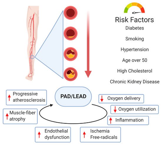
LEAD usually involves atherosclerotic disease in the abdominal aorta, iliac, and femoral arteries. The pathophysiology of atherosclerotic plaque involves complex interactions between cholesterol metabolism and vascular cell activity. It is also known that disturbance of the laminar arterial flow in PAD plays an essential role in the adhesion of inflammatory blood cells to the arterial wall and plays a role in plaque generation [14]. The hemodynamic consequences of atherosclerotic plaque depend on the degree of stenosis/occlusion. A narrowed vessel progresses towards chronic total occlusion. LEAD patients demonstrate high grades of inflammation and active oxidative stress, which are vital mechanisms in PAD pathophysiology [14]. There is an interest in the vaso-protective effects of heme oxygenase 1 (HO-1) as a potential antioxidant by affecting the proliferation, migration, and adhesion of smooth vascular muscle cells, endothelial cells, and leukocytes [15]. It was previously suggested that PE acts on endothelial functions by regulating genes involved in modulating oxidative metabolism, cell apoptosis, cell growth and proliferation, and endothelial vascular nitric oxide synthase (eNOS) [14][16].
Vessel wall remodelling and angiogenesis in peripheral arteries appear to be the most important tissue response process to the atherosclerotic injuries [17]. Extracellular matrix (ECM) provides a mechanical scaffold and support to cell migration regulated by cytokines, growth factors, and enzymes, such as matrix metalloproteinases (MMPs) [18][19]. Hernandez-Aguilera et al. suggested that atherosclerosis of the lower extremities is due to an excessive reparative response, which includes factors favoring ECM degradation [19][20]. Decreased levels of structural proteins of the arterial wall are highly influenced by MMP activity [19]. These proteins also have defined roles in maintaining normal function and migration of smooth muscle cells [21]. Connective tissue turnover in the structural and signaling properties of the arterial cells plays a central role in the LEAD development [22]. Single nucleotide polymorphisms of the genes encoding for some MMPs have also been associated with the risk of developing LEAD. Blankenberg et al. [23] reported a study that focused on two MMP-9 gene polymorphisms: the MMP-9/C-1562T promoter polymorphism, which influences the transcriptional activity of the MMP-9 gene, and the exonic MMP-9/R279Q polymorphism, which leads to an amino acid exchange in the catalytic domain of MMP-9. The authors described a significant association between the R279Q polymorphism and cardiovascular events in patients with stable angina.
Arterial calcification was previously considered a passive degenerative process but is now recognized as a complex process actively regulated by several cell molecules [24]. Arterial calcification mainly involves the intima and media, and is associated with cardiovascular risk factors, such as diabetes, hypertension, hyperlipidemia, and chronic kidney disease [25]. Arterial calcification is related to the apoptosis of vascular smooth muscle cells and macrophages [26]. Studies provided by Hui laboratory [27] focused on the genetic variants of the matrix Gla protein (MGP), a protein known as a key player in in vivo inhibition of calcification. The authors analyzed rs4236, rs1800801, and rs1800802 variants of the MGP gene, and showed association with calcification on the arterial wall but not with calcification in atherosclerotic plaques [27]. Other studies showed that different types of arterial calcification develop through various molecular mechanisms in different vessel types [28]. In contrast to carotid and coronary arteries, arterial calcification of LEAD is mostly located in the media.
Several genotypes serve as potential risk factors for atherosclerosis. However, evidence of their clinical relevance is weak. Some of the most common risk factors for PAD (diabetes, dyslipidemia) are heritable. However, PAD may also result from genetic factors acting independently. Identification of such genes may provide insights into pathophysiologic mechanisms of PAD progress and facilitate the development of novel therapeutic approaches [29]. In contrast to coronary artery disease, relatively few genetic variants that influence susceptibility to PAD have been discovered because there may be more significant clinical and genetic heterogeneity in PAD patients. Definitively, genetic factors may have an impact on the early onset of PAD in young adults, including these mechanisms affecting the process of inflammation, thrombosis, and the metabolism of cholesterol and homocysteine. Among these genetic disorders, familial hypercholesterolemia or hyperhomocysteinemia are the most known. For example, Flex et al. [30] explored the association between the interleukin-6 gene (IL-6)-174 G/C single nucleotide polymorphism (SNP) and the risk of peripheral artery occlusive disease. The authors concluded that the analyzed polymorphism is important in the pathophysiology of ischemic diseases of the lower extremity [30]. Several other candidate genes involved in the process of atherosclerosis and regulating inflammation and coagulation pathways and vascular matrix regulation were also analyzed, such as β-fibrinogen, eNOS, MTHFR, and glutathione S-transferase [31]. However, any reported associations between variants in these genes and PAD have not been confirmed [29].

3.1. Neovascularization

In healthy individuals, reducing blood flow to the lower extremity is followed by a physiological process to limit ischemia in the distal tissue by expanding the vasculature. Neovascularization is regulated by mechanisms that respond to ischemia, hypoxia, or shear stress [32]. Neovascularization is the process of blood vessel development and growth through three distinct biological processes: vasculogenesis, angiogenesis, and arteriogenesis [4]. Two forms of neovascularization, angiogenesis, and arteriogenesis can be distinguished during LEAD. Physiological studies supported the hypothesis that after ischemia, the neovascularization process can re-establish blood flow through the ischemic tissue and protect the extremities from blood flow loss. It is important to note that compromised arterial inflow results in angiogenesis due to distal tissue ischemia and arteriogenesis that occurs close to the site of arterial trunk occlusion [32]. Structural and functional adaptations of the vasculature can also be induced by exercise training in humans [33]. LEAD symptoms may be the result of not only an obstructive vascular process but also an impaired neovascularization response [1].

3.2. Vasculogenesis

During embryonal development, blood vessels first appear due to vasculogenesis, the formation of capillaries from endothelial cells differentiating in situ from groups of mesodermal cells [2]. During embryonal life, microvascular collaterals develop after the arterial trees. These collaterals increase their diameter and wall thickness as a consequence of the growth of a muscular layer [4][34]. Hemodynamic forces regulate the differentiation of arteries and veins from the primary vascular plexus. It was suggested that vessels adapt to flow during arteriogenesis [4]. Other observations have indicated that vasculogenesis is not limited to early embryogenesis but also has a physiological role during vascular disease in adults. Postnatal vasculogenesis was explained by the presence of circulating endothelial cells and endothelial progenitor cells in human and several mechanisms of blood vessel formation during tumor growth [4][35].

3.3. Angiogenesis

Angiogenesis is the process of new capillary formation from pre-existing capillary beds that involves proliferation, sprouting, and migration of endothelial cell migration. Angiogenesis occurs naturally during wound healing, tissue growth, and repair. The process of angiogenesis is highly controlled, dependent on a balance of both pro-angiogenic and anti-angiogenic factors. The process of angiogenesis results from complex interactions between growth factors, endothelial cells, pericytes, fibroblasts, smooth muscle cells, and the extracellular matrix. As a result of these interactions, extracellular proteolysis, endothelial cell migration, proliferation, and differentiation, and finally, vascular wall remodeling can occur [4]. Vascular endothelial growth factor (VEGF) is the main factor stimulating angiogenesis in response to tissue hypoxia [36]. Angiogenesis is primarily stimulated by tissue hypoxia via activation of hypoxia inducible factor (HIF). Specifically, in the absence of cellular oxygen, hypoxia-inducible factors (HIFs) are activated and translocated to the nucleus where they upregulate genes including VEGFA, angiopoietins, and nitric oxide [1][2][37]. Through signaling by VEGF, mature endothelial cells (ECs) are directed to sites of hypoxia to participate in the formation of new blood vessels. The endothelial cell activation is associated with cytokine release, initiation of vasodilation, and increased endothelial cell permeability. VEGF promotes the release of many proteolytic factors, such as matrix metalloproteases, which degrade the extracellular matrix and facilitate endothelial cell migration [38]. Once a functional vascular network is formed, the new vessels are remodeled to become a mature vessel system [32]. Angiogenesis describes the vascular tissue remodeling and is characterized by the expansion of existing capillary density to reach ischemic sites, maintaining the lower extremity function in patients with LEAD.

3.4. Arteriogenesis

As a distinct process from angiogenesis, arteriogenesis refers to the growth of new arteries and arterioles either de novo or from pre-existing arterial collaterals [3][39]. It mainly involves the proliferation of vascular endothelial cells (ECs) and smooth muscle cells (SMCs) [40]. As a result of the occlusion of the main arterial trunk, the pre-existing arterio-arteriolar anastomoses between interconnected perfusion territories can undergo adaptive enlargement that develops into a functional network of collateral arteries [32]. Arteriogenesis is critical to the restoration of tissue perfusion following the development of a functionally significant decrease of arterial inflow during LEAD [41]. The transformation of native microvascular collateral arterioles into functional arteries with consequent recovery of blood flow is particularly observed after ischemic injury [42].
It was previously suggested that vessel segments could adapt to the amount of flow [43]. Collateral growth is driven by hemodynamic forces and leads to initial vasodilation due to increased levels of nitric oxide [44]. While hypoxia is the primary driver of angiogenesis, arteriogenesis is mainly induced by a combination of shear stress and other mechanical factors [36][39][41]. Pulsatile shear stress activates the cascade of events that leads to the development of collateral circulation [4][45]. Several genes are controlled by shear stress-responsive elements in their promotor, and fluid shear stress influences these genes’ expression. The role of mechano-sensors and transducers that convey the shear stress message during collateral remodeling has been suggested as a mechanism directing neovascularization [46][47][48]. A fluid shear stress-associated transient receptor potential cation channel, subfamily V, member 4 (trpv4), turned out to be upregulated transiently after endurance training [49][50]. Shear stress increases the expression of an isoform of connexin, connexin-37, in endothelial cells [51].
Arteriogenesis consist of two phases: the early inflammation phase and the later phase of vessel diameter increase, remodeling, and maturation. Hemodynamic forces in the collateral vessels are the primary stimulus to initiate arteriogenesis. As a result of the increased shear stress force, endothelial cells express monocyte adhesion molecules: platelet endothelial cell adhesion molecule (PECAM-1), MCP-1, intracellular adhesion molecule (ICAM-1), and vascular cell adhesion molecule (VCAM-1) [52]. Endothelial cells regulate adhesion molecule gene expression through a mechano-transduction process by specific shear stress receptors [32]. Cytokines and cell adhesion molecules attract monocytes to adhere and invade the vascular wall. Recruited monocytes infiltrate the vessel wall and transform into macrophages. Once activated, they produce TNF-α and attract more monocytes. Recruitment of circulating monocytes and resident macrophages promotes arteriogenesis by their ability to secrete metalloproteinases, chemokines, and growth factors [53]. The proliferation of the endothelium is followed by smooth muscle proliferation and migration to form a new neointima. Little is known about the mechanisms triggering SMC proliferation in arteriogenesis. Growth factors, such as fibroblast growth factor 2 (FGF-2) and platelet-derived growth factor-BB (PDGF-BB), are essential for SMC proliferation in arteriogenesis [54]. Finally, enlargement of the blood vessel occurs by remodeling the adventitia through fibroblast activation and proliferation. These events lead to new collateral vessel development in postnatal life.
Arteriogenesis differs from angiogenesis in several aspects, the most important being the dependence of angiogenesis on hypoxia and the dependence of arteriogenesis on inflammation. Arteriogenesis occurs in tissues near arterial stenosis/occlusion, whereas peripheral ischemic regions undergo angiogenesis, which is the growth of new capillaries [55]. Collateral vessels resulting from arteriogenesis are typically surrounded by normoxic tissue [56]. While molecular regulation of angiogenesis is well analyzed, events regulating arteriogenesis are still controversial. As well as for angiogenesis, VEGF is also critical to arteriogenesis [57]. In arteriogenesis, VEGF activation of extracellular signal-regulating kinase 1/2 (ERK1/2) induces endothelial cell proliferation, network formation, and increased lumen size. Disruption of VEGF-induced endothelial ERK1/2 signaling results in decreased arteriogenesis [58]. The activation of this signaling is modulated by NRP-1 [59]. VEGF, TGF-α, and FGF-2 stimulate the proliferation of endothelial cells, whereas TGF-β, GM-CSF, monocyte chemoattractant protein-1 (MCP-1), and PDGF stimulate arteriogenesis through the proliferation of smooth muscle cells [60]. The proliferation of endothelial cells and smooth muscle cells leads to lumen size expansion of the collateral artery [61]. In contrast to angiogenesis, arteriogenesis requires a coordinated response that involves multiple cell types, not only endothelial and vascular smooth muscle cells [41]. Several inflammatory cells, including lymphocytes, natural killer (NK) cells, macrophages, and mast cells, were also suggested to play a role in arteriogenesis [62][63][64]. The presence of inflammatory cells is critical as these cells serve as the major source of VEGF in the absence of tissue ischemia [41][65][66]. Mast cells have also been associated with arteriogenesis and collateral formation [67][68]. The proinflammatory response during arteriogenesis is beneficial in restoring blood flow, but may lead to enhanced progression of atherosclerosis [68].
Collateral circulation occurs between arterioles in most healthy tissues and in pathological conditions like ischemic injury [39]. Arteriogenesis can be induced by regular exercise or muscle loading in humans in physiological conditions [33][49][69]. A more regular exercise regimen was also proposed to be required for sufficient arteriogenesis to compensate for an arterial occlusion in LEAD [49]. It explains why significant stenosis/occlusion of main arteries may remain asymptomatic in patients with chronic LEAD for many years. Exercise therapy can influence the vascular system supply to the ischemic limb by promoting both angiogenesis and arteriogenesis [33]. An increase of angiogenic and arteriogenic factors, such as VEGF and HIF-1 alpha, was observed after regular exercise [70]. Additionally, higher concentrations of heat shock proteins and the enzyme nitric oxide synthase (NOS) in blood during exercise therapy was noticed [71]. Nitric oxide (NO) is an important cellular signaling molecule produced in high levels in muscle by neuronal NOS [33]. Under ischemic conditions, the role of NO in vasodilatation is increased compared with non-ischemic conditions, which results in an up-regulation of endothelial NOS (eNOS) [72]. Regular exercise training in the patient with LEAD leads to similar mechanisms of vascular adaptation such as increased fluid shear stress and promotes arteriogenesis. After successful revascularization, collateral arteries shrink or disappear as the main blood flow is directed back through the re-opened artery or vascular reconstruction. Supervised exercise therapy is considered to be a potential therapeutic option in chronic LEAD patients under conservative treatment.

References

  1. Bonham, C.A.; Kuehlmann, B.; Gurtner, G.C. Impaired Neovascularization in Aging. Adv. Wound Care 2020, 9, 111–126.
  2. Carmeliet, P. Mechanisms of angiogenesis and lymphangiogenesis. Nat. Med. 2000, 6, 389–395.
  3. Helisch, A.; Schaper, W. Arteriogenesis: The development and growth of collateral arteries. Microcirculation 2003, 10, 83–97.
  4. Rizzi, A.; Benagiano, V.; Ribatti, D. Angiogenesis versus arteriogenesis. Rom. J. Morphol. Embryol. 2017, 58, 15–19.
  5. Fowkes, F.G.R.; Rudan, D.; Rudan, I.; Aboyans, V.; Denenberg, J.O.; McDermott, M.M.; Norman, P.E.; Sampson, U.K.A.; Williams, L.J.; Mensah, G.A.; et al. Comparison of global estimates of prevalence and risk factors for peripheral artery disease in 2000 and 2010: A systematic review and analysis. Lancet 2013, 382, 1329–1340.
  6. Sartipy, F.; Sigvant, B.; Lundin, F.; Wahlberg, E. Ten Year Mortality in Different Peripheral Arterial Disease Stages: A Population Based Observational Study on Outcome. Eur. J. Vasc. Endovasc. Surg. 2018, 55, 529–536.
  7. Aboyans, V.; Ricco, J.B.; Bartelink, M.L.E.L.; Björck, M.; Brodmann, M.; Cohnert, T.; Collet, J.P.; Czerny, M.; De Carlo, M.; Debus, S.; et al. 2017 ESC Guidelines on the Diagnosis and Treatment of Peripheral Arterial Diseases, in collaboration with the European Society for Vascular Surgery (ESVS). Eur. Heart J. 2018, 39, 763–816.
  8. Norgren, L.; Hiatt, W.R.; Dormandy, J.A.; Nehler, M.R.; Harris, K.A.; Fowkes, F.G.R. Inter-Society Consensus for the Management of Peripheral Arterial Disease (TASC II). J. Vasc. Surg. 2007, 45, S5–S67.
  9. De Weerd, M.; Greving, J.P.; De Jong, A.W.F.; Buskens, E.; Bots, M.L. Prevalence of asymptomatic carotid artery stenosis according to age and sex systematic review and metaregression analysis. Stroke 2009, 40, 1105–1113.
  10. Criqui, M.H.; Aboyans, V. Epidemiology of Peripheral Artery Disease. Circ. Res. 2015, 116, 1509–1526.
  11. Jude, E.B.; Oyibo, S.O.; Chalmers, N.; Boulton, A.J.M. Peripheral arterial disease in diabetic and nondiabetic patients: A comparison of severity and outcome. Diabetes Care 2001, 24, 1433–1437.
  12. Aboyans, V.; Criqui, M.H.; Denenberg, J.O.; Knoke, J.D.; Ridker, P.M.; Fronek, A. Risk factors for progression of peripheral arterial disease in large and small vessels. Circulation 2006, 113, 2623–2629.
  13. Stone, P.A.; Yacoub, M. Inflammatory biomarkers in peripheral arterial disease. Semin. Vasc. Surg. 2014, 27, 148–151.
  14. Signorelli, S.S.; Marino, E.; Scuto, S.; Di Raimondo, D. Pathophysiology of peripheral arterial disease (Pad): A review on oxidative disorders. Int. J. Mol. Sci. 2020, 21, 4393.
  15. Kishimoto, Y.; Ibe, S.; Saita, E.; Sasaki, K.; Niki, H.; Miura, K.; Ikegami, Y.; Ohmori, R.; Kondo, K.; Momiyama, Y. Plasma Heme Oxygenase-1 Levels in Patients with Coronary and Peripheral Artery Diseases. Dis. Markers 2018, 2018.
  16. Walker, M.A.; Hoier, B.; Walker, P.J.; Schulze, K.; Bangsbo, J.; Hellsten, Y.; Askew, C.D. Vasoactive enzymes and blood flow responses to passive and active exercise in peripheral arterial disease. Atherosclerosis 2016, 246, 98–105.
  17. Xu, J.; Shi, G.P. Vascular wall extracellular matrix proteins and vascular diseases. Biochim. Biophys. Acta Mol. Basis Dis. 2014, 1842, 2106–2119.
  18. Tanaka, L.Y.; Laurindo, F.R.M. Vascular remodeling: A redox-modulated mechanism of vessel caliber regulation. Free Radic. Biol. Med. 2017, 109, 11–21.
  19. Hernández-Aguilera, A.; Nielsen, S.H.; Bonache, C.; Fernández-Arroyo, S.; Martín-Paredero, V.; Fibla, M.; Karsdal, M.A.; Genovese, F.; Menendez, J.A.; Camps, J.; et al. Assessment of extracellular matrix-related biomarkers in patients with lower extremity artery disease. J. Vasc. Surg. 2018, 68, 1135–1142.e6.
  20. Bentzon, J.F.; Otsuka, F.; Virmani, R.; Falk, E. Mechanisms of Plaque Formation and Rupture. Circ. Res. 2014, 114, 1852–1866.
  21. Wight, T.N.; Merrilees, M.J. Proteoglycans in atherosclerosis and restenosis: Key roles for versican. Circ. Res. 2004, 94, 1158–1167.
  22. Vassiliadis, E.; Barascuk, N.; Karsdal, M.A. Atherofibrosis—A unique and common process of the disease pathogenesis of atherosclerosis and fibrosis—Lessons for biomarker development. Am. J. Transl. Res. 2013, 5, 1–14.
  23. Blankenberg, S.; Rupprecht, H.J.; Poirier, O.; Bickel, C.; Smieja, M.; Hafner, G.; Meyer, J.; Cambien, F.; Tiret, L. Plasma concentrations and genetic variation of matrix metalloproteinase 9 and prognosis of patients with cardiovascular disease. Circulation 2003, 107, 1579–1585.
  24. Yan, H.; Chang, Z.; Liu, Z. The risk factors for calcification vary among the different sections of the lower extremity artery in patients with symptomatic peripheral arterial disease. BMC Cardiovasc. Disord. 2020, 20.
  25. Demer, L.L.; Tintut, Y. Inflammatory, metabolic, and genetic mechanisms of vascular calcification. Arterioscler. Thromb. Vasc. Biol. 2014, 34, 715–723.
  26. Chistiakov, D.A.; Myasoedova, V.A.; Melnichenko, A.A.; Grechko, A.V.; Orekhov, A.N. Calcifying Matrix Vesicles and Atherosclerosis. BioMed Res. Int. 2017, 2017.
  27. Wang, Y.; Chen, J.; Zhang, Y.; Yu, W.; Zhang, C.; Gong, L.; Shao, L.; Lu, J.; Gao, Y.; Chen, X.; et al. Common genetic variants of MGP are associated with calcification on the arterial wall but not with calcification present in the atherosclerotic plaques. Circ. Cardiovasc. Genet. 2013, 6, 271–278.
  28. Lanzer, P.; Boehm, M.; Sorribas, V.; Thiriet, M.; Janzen, J.; Zeller, T.; St Hilaire, C.; Shanahan, C. Medial vascular calcification revisited: Review and perspectives. Eur. Heart J. 2014, 35, 1515–1525.
  29. Kullo, I.J.; Leeper, N.J. The Genetic Basis of Peripheral Arterial Disease: Current Knowledge, Challenges, and Future Directions. Circ. Res. 2015, 116, 1551–1560.
  30. Flex, A.; Gaetani, E.; Pola, R.; Santoliquido, A.; Aloi, F.; Papaleo, P.; Dal Lago, A.; Pola, E.; Serricchio, M.; Tondi, P.; et al. The -174 G/C polymorphism of the interleukin-6 gene promoter is associated with peripheral artery occlusive disease. Eur. J. Vasc. Endovasc. Surg. 2002, 24, 264–268.
  31. Li, R.; Folsom, A.R.; Sharrett, A.R.; Couper, D.; Bray, M.; Tyroler, H.A. Interaction of the glutathione S-transferase genes and cigarette smoking on risk of lower extremity arterial disease: The Atherosclerosis Risk in Communities (ARIC) study. Atherosclerosis 2001, 154, 729–738.
  32. Arellano Mendoza, M.; Robles, H.; Romo, E.; Rios, A.; Escalante, B. Nitric Oxide-Dependent Neovascularization Role in the Lower Extremity Disease. Curr. Pharm. Des. 2007, 13, 3591–3596.
  33. Vogel, J.; Niederer, D.; Jung, G.; Troidl, K. Exercise-Induced Vascular Adaptations under Artificially Versus Pathologically Reduced Blood Flow: A Focus Review with Special Emphasis on Arteriogenesis. Cells 2020, 9, 333.
  34. Chalothorn, D.; Faber, J.E. Formation and maturation of the native cerebral collateral circulation. J. Mol. Cell. Cardiol. 2010, 49, 251–259.
  35. Ribatti, D.; Vacca, A.; Nico, B.; Roncali, L.; Dammacco, F. Postnatal vasculogenesis. Mech. Dev. 2001, 100, 157–163.
  36. Simons, M.; Eichmann, A. Molecular controls of arterial morphogenesis. Circ. Res. 2015, 116, 1712–1724.
  37. Pugh, C.W.; Ratcliffe, P.J. Regulation of angiogenesis by hypoxia: Role of the HIF system. Nat. Med. 2003, 9, 677–684.
  38. Stetler-Stevenson, W.G. Matrix metalloproteinases in angiogenesis: A moving target for therapeutic intervention. J. Clin. Investig. 1999, 103, 1237–1241.
  39. Faber, J.E.; Chilian, W.M.; Deindl, E.; Van Royen, N.; Simons, M. A brief etymology of the collateral circulation. Arterioscler. Thromb. Vasc. Biol. 2014, 34, 1854–1859.
  40. Lasch, M.; Caballero Martinez, A.; Kumaraswami, K.; Ishikawa-Ankerhold, H.; Meister, S.; Deindl, E. Contribution of the Potassium Channels KV1.3 and KCa3.1 to Smooth Muscle Cell Proliferation in Growing Collateral Arteries. Cells 2020, 9, 913.
  41. Ricard, N.; Zhang, J.; Zhuang, Z.W.; Simons, M. Isoform-Specific Roles of ERK1 and ERK2 in Arteriogenesis. Cells 2019, 9, 38.
  42. Sealock, R.; Zhang, H.; Lucitti, J.L.; Moore, S.M.; Faber, J.E. Congenic fine-mapping identifies a major causal locus for variation in the native collateral circulation and ischemic injury in brain and lower extremity. Circ. Res. 2014, 114, 660–671.
  43. Peirce, S.M.; Skalak, T.C. Microvascular remodeling: A complex continuum spanning angiogenesis to arteriogenesis. Microcirculation 2003, 10, 99–111.
  44. Troidl, K.; Tribulova, S.; Cai, W.J.; Rüding, I.; Apfelbeck, H.; Schierling, W.; Troidl, C.; Schmitz-Rixen, T.; Schaper, W. Effects of endogenous nitric oxide and of deta nonoate in arteriogenesis. J. Cardiovasc. Pharmacol. 2010, 55, 153–160.
  45. Buschmann, I.; Pries, A.; Styp-Rekowska, B.; Hillmeister, P.; Loufrani, L.; Henrion, D.; Shi, Y.; Duelsner, A.; Hoefer, I.; Gatzke, N.; et al. Pulsatile shear and Gja5 modulate arterial identity and remodeling events during flow-driven arteriogenesis. Development 2010, 137, 2187–2196.
  46. Tronc, F.; Mallat, Z.; Lehoux, S.; Wassef, M.; Esposito, B.; Tedgui, A. Role of Matrix Metalloproteinases in Blood Flow–Induced Arterial Enlargement. Arterioscler. Thromb. Vasc. Biol. 2000, 20.
  47. Shi, Z.D.; Tarbell, J.M. Fluid flow mechanotransduction in vascular smooth muscle cells and fibroblasts. Ann. Biomed. Eng. 2011, 39, 1608–1619.
  48. Gerhold, K.A.; Schwartz, M.A. Ion channels in endothelial responses to fluid shear stress. Physiology 2016, 31, 359–369.
  49. Sayed, A.; Schierling, W.; Troidl, K.; Rüding, I.; Nelson, K.; Apfelbeck, H.; Benli, I.; Schaper, W.; Schmitz-Rixen, T. Exercise linked to transient increase in expression and activity of cation channels in newly formed hind-limb collaterals. Eur. J. Vasc. Endovasc. Surg. 2010, 40, 81–87.
  50. Troidl, C.; Troidl, K.; Schierling, W.; Cai, W.J.; Nef, H.; Möllmann, H.; Kostin, S.; Schimanski, S.; Hammer, L.; Elsässer, A.; et al. Trpv4 induces collateral vessel growth during regeneration of the arterial circulation. J. Cell. Mol. Med. 2009, 13, 2613–2621.
  51. Pfenniger, A.; Wong, C.; Sutter, E.; Cuhlmann, S.; Dunoyer-Geindre, S.; Mach, F.; Horrevoets, A.J.; Evans, P.C.; Krams, R.; Kwak, B.R. Shear stress modulates the expression of the atheroprotective protein Cx37 in endothelial cells. J. Mol. Cell. Cardiol. 2012, 53, 299–309.
  52. Chen, Z.; Rubin, J.; Tzima, E. Role of PECAM-1 in arteriogenesis and specification of preexisting collaterals. Circ. Res. 2010, 107, 1355–1363.
  53. Takeda, Y.; Costa, S.; Delamarre, E.; Roncal, C.; Leite De Oliveira, R.; Squadrito, M.L.; Finisguerra, V.; Deschoemaeker, S.; Bruyère, F.; Wenes, M.; et al. Macrophage skewing by Phd2 haplodeficiency prevents ischaemia by inducing arteriogenesis. Nature 2011, 479, 122–126.
  54. de Paula, E.V.; Flores-Nascimento, M.C.; Arruda, V.R.; Garcia, R.A.; Ramos, C.D.; Guillaumon, A.T.; Annichino-Bizzacchi, J.M. Dual gene transfer of fibroblast growth factor-2 and platelet derived growth factor-BB using plasmid deoxyribonucleic acid promotes effective angiogenesis and arteriogenesis in a rodent model of hindlimb ischemia. Transl. Res. 2009, 153, 232–239.
  55. Troidl, K.; Schubert, C.; Vlacil, A.K.; Chennupati, R.; Koch, S.; Schütt, J.; Oberoi, R.; Schaper, W.; Schmitz-Rixen, T.; Schieffer, B.; et al. The Lipopeptide MALP-2 Promotes Collateral Growth. Cells 2020, 9, 997.
  56. Deindl, E.; Buschmann, I.; Hoefer, I.E.; Podzuweit, T.; Boengler, K.; Vogel, S.; Van Royen, N.; Fernandez, B.; Schaper, W. Role of ischemia and of hypoxia-inducible genes in arteriogenesis after femoral artery occlusion in the rabbit. Circ. Res. 2001, 89, 779–786.
  57. Moraes, F.; Paye, J.; Gabhann, F.M.; Zhuang, Z.W.; Zhang, J.; Lanahan, A.A.; Simons, M. Endothelial cell-dependent regulation of arteriogenesis. Circ. Res. 2013, 113, 1076–1086.
  58. Lanahan, A.A.; Lech, D.; Dubrac, A.; Zhang, J.; Zhuang, Z.W.; Eichmann, A.; Simons, M. PTP1b is a physiologic regulator of vascular endothelial growth factor signaling in endothelial cells. Circulation 2014, 130, 902–909.
  59. Lanahan, A.; Zhang, X.; Fantin, A.; Zhuang, Z.; Rivera-Molina, F.; Speichinger, K.; Prahst, C.; Zhang, J.; Wang, Y.; Davis, G.; et al. The Neuropilin 1 Cytoplasmic Domain Is Required for VEGF-A-Dependent Arteriogenesis. Dev. Cell 2013, 25, 156–168.
  60. Van Royen, N.; Piek, J.J.; Buschmann, I.; Hoefer, I.; Voskuil, M.; Schaper, W. Stimulation of arteriogenesis; a new concept for the treatment of arterial occlusive disease. Cardiovasc. Res. 2001, 49, 543–553.
  61. Shireman, P.K. The chemokine system in arteriogenesis and hind limb ischemia. J. Vasc. Surg. 2007, 45, A48–A56.
  62. Stabile, E.; Kinnaird, T.; La Sala, A.; Hanson, S.K.; Watkins, C.; Campia, U.; Shou, M.; Zbinden, S.; Fuchs, S.; Kornfeld, H.; et al. CD8+ T lymphocytes regulate the arteriogenic response to ischemia by infiltrating the site of collateral vessel development and recruiting CD4+ mononuclear cells through the expression of interleukin-16. Circulation 2006, 113, 118–124.
  63. Krishnasamy, K.; Limbourg, A.; Kapanadze, T.; Gamrekelashvili, J.; Beger, C.; Häger, C.; Lozanovski, V.J.; Falk, C.S.; Napp, L.C.; Bauersachs, J.; et al. Blood vessel control of macrophage maturation promotes arteriogenesis in ischemia. Nat. Commun. 2017, 8, 1–14.
  64. Ribatti, D. A new role of mast cells in arteriogenesis. Microvasc. Res. 2018, 118, 57–60.
  65. Heil, M.; Ziegelhoeffer, T.; Pipp, F.; Kostin, S.; Martin, S.; Clauss, M.; Schaper, W. Blood monocyte concentration is critical for enhancement of collateral artery growth. Am. J. Physiol. Hear. Circ. Physiol. 2002, 283.
  66. Morrison, A.R.; Yarovinsky, T.O.; Young, B.D.; Moraes, F.; Ross, T.D.; Ceneri, N.; Zhang, J.; Zhuang, Z.W.; Sinusas, A.J.; Pardi, R.; et al. Chemokine-coupled β2 integrin-induced macrophage Rac2-myosin IIA interaction regulates VEGF-A mRNA stability and arteriogenesis. J. Exp. Med. 2014, 211, 1957–1968.
  67. Chillo, O.; Kleinert, E.C.; Lautz, T.; Lasch, M.; Pagel, J.I.; Heun, Y.; Troidl, K.; Fischer, S.; Caballero-Martinez, A.; Mauer, A.; et al. Perivascular Mast Cells Govern Shear Stress-Induced Arteriogenesis by Orchestrating Leukocyte Function. Cell Rep. 2016, 16, 2197–2207.
  68. Bot, I.; van der Velden, D.; Bouwman, M.; Kröner, M.J.; Kuiper, J.; Quax, P.H.A.; de Vries, M.R. Local Mast Cell Activation Promotes Neovascularization. Cells 2020, 9, 701.
  69. Dopheide, J.F.; Rubrech, J.; Trumpp, A.; Geissler, P.; Zeller, G.C.; Schnorbus, B.; Schmidt, F.; Gori, T.; Münzel, T.; Espinola-Klein, C. Supervised exercise training in peripheral arterial disease increases vascular shear stress and profunda femoral artery diameter. Eur. J. Prev. Cardiol. 2017, 24, 178–191.
  70. Pope, Z.K.; Willardson, J.M.; Schoenfeld, B.J. Exercise and blood flow restriction. J. Strength Cond. Res. 2013, 27, 2914–2926.
  71. Loenneke, J.P.; Wilson, G.J.; Wilson, J.M. A mechanistic approach to blood flow occlusion. Int. J. Sports Med. 2010, 31, 1–4.
  72. Casey, D.P.; Madery, B.D.; Curry, T.B.; Eisenach, J.H.; Wilkins, B.W.; Joyner, M.J. Nitric oxide contributes to the augmented vasodilatation during hypoxic exercise. J. Physiol. 2010, 588, 373–385.
More
Information
Subjects: Pathology
Contributor MDPI registered users' name will be linked to their SciProfiles pages. To register with us, please refer to https://encyclopedia.pub/register :
View Times: 401
Revisions: 2 times (View History)
Update Date: 24 May 2021
1000/1000