1. Vertebrata
All multicellular organisms, vertebrates or invertebrates, possess an immune system that is an essential component of the defense strategies to recognize and neutralize parasites, microorganisms, viruses, and more. Vertebrates have two lines of defense, defined as innate, or non-specific, immunity and adaptive, or acquired, immunity. The success of the immune response is guaranteed by a complex interweaving of interactions between different types of molecules, each with specific functions.
In mammals, the expression of
AIF-1 has been reported in different cell types such as activated macrophages, microglial cells, and dendritic cells (DC). The main immunomodulatory role of the protein during the inflammatory response has also been highlighted in these cells. However, this gene plays different roles in the nervous and immune systems
[1][2]. Furthermore,
AIF-1 is also expressed in muscle, liver, spleen, and thymus in rats
[3] and humans
[4], and it is considered a marker of activated human vascular smooth muscle cells and arterial injury
[5]. A possible link between skeletal muscle cell proliferation and AIF-1-induced inhibition of satellite cell proliferation has also been revealed
[4], expanding the possible fields of action of this protein. In 2017, Elizondo et al. underlined the importance of AIF-1 in antigen presentation by DC
[6]. In this study, they reported that
AIF-1 is expressed in CD11c
+ dendritic cells and that expression silencing restrains induction of antigen-specific CD4
+ T cell effector responses. Moreover, because
AIF-1 knockdown in murine DC resulted in impaired T cell proliferation, the same authors demonstrated that
AIF-1 expression in DC serves as a potent governor of cognate T cell responses
[7]. Furthermore, Miyata et al.
[8] showed an upregulation of
AIF-1 transcripts in red seabream (Teleostean) leukocytes upon LPS stimulation and suggested a similar function in Vertebrata. More recently, a novel role of AIF-1 as a Ca
2+-responsive scaffold protein involved in cell differentiation emerged
[9]. In particular, the requirement of
AIF-1 expression in hematopoietic progenitors for differentiation into Mo-DC and cDC1 subsets has been evidenced.
The AIF-1 protein represents a crucial element for macrophages’ survival and pro-inflammatory activity
[10][11]. It is involved in inflammation and immune responses associated with autoimmune diseases
[12], and also with vasculopathy
[13] and CNS injury
[14]. In a recent review, Sikora et al.
[15] summarized the role of AIF-1 in the pathogenesis of some diseases including endometriosis, breast cancer, atherosclerosis, rheumatoid arthritis, and fibrosis. Indeed, several authors evidenced its importance in rheumatoid arthritis progression
[16][17][18].
AIF-1 is also considered as a new risk factor for the development of atherosclerosis
[19], and, when overexpressed, it influences the intensification of atherosclerotic plaque calcification
[20]. Recent studies report a tight link between AIF-1 and cancer. It is involved in breast cancer development by interacting with several proteins such as metalloproteinases
[21] or transcription factors
[22] and activating the downstream pathways, inducing cell proliferation. Interestingly,
AIF-1 is also expressed in the CNS of vertebrates. Mostly known under the name of Iba1, this factor is largely recognized as a specific microglial marker allowing the distinction of these brain-resident immune cells from neurons and other brain glial cells
[23].
AIF-1 is modulated upon different brain injuries and pathologies, indicating a link with CNS inflammatory states
[24].
AIF-1 expression is generally linked to the presence of activated brain microglia/macrophages. Beschorner et al.
[25] reported the expression of
AIF-1 in a limited subpopulation of microglial cells and did not observe a significant upregulation of this gene upon traumatic brain injury. In contrast, Schwab et al.
[26] observed an important accumulation of AIF-1
+ cells in microglia/macrophages in association with experimental spinal cord injury. Indeed, the authors indicate this accumulation as essential for the initiation of an effective response to CNS injury and repair events. Interestingly,
AIF-1 expression has been reported in human microglia following cerebral infarction
[27]. This suggests that
AIF-1 can also be upregulated by non-inflammatory brain lesions such as hypoxia.
2. Invertebrata
Despite the limited amount of published data, it appears that in vertebrates, like in vertebrates, AIF-1 is mainly involved in the inflammatory response. To date, AIF-1 genes have been characterized functionally in phylogenetically distant group of species, such as sponges, cnidarians, mollusks, annelids, and echinoderms. Many invertebrate species are of commercial interest, and more and more research groups are directing their investigations to these animals and their defense mechanisms. Aquaculture of numerous species suffers from massive infections and severe mortality. Therefore, it is necessary to understand and solve disease-related problems. Furthermore, due to the relative simplicity of their immune system, invertebrates also represent good models for studying innate immunity and providing insight into the evolution of their defense mechanisms.
Invertebrates are characterized by a lack of acquired immunity; thus, in such organisms the innate immune systems can provide the host with an immediate defense against pathogens in a non-specific manner. During this event, the immune system is able to differentiate self from non-self. The recognition process is carried out by circulating cells named amebocytes, hemocytes, or coelomocytes according to the various animal groups. In many invertebrate species, these cells have a macrophage-like appearance and function. They are characterized by the presence on their surface of pathogen recognition receptors (PRRs) recognizing pathogen-associated molecular patterns (PAMPs). These are well-conserved molecular structures expressed by various pathogens whether they are viruses, bacteria, or other foreign particles. The receptor–ligand binding triggers a complex cascade of cellular reactions with the production of a wide array of effector molecules.
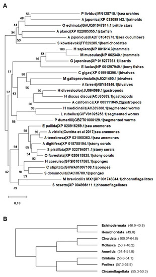
AIF-1 is a ubiquitously expressed and well-conserved molecule involved in innate immunity response from sponges
[28][29] to humans
[30]. Notably, when compared all together, AIF-1-related proteins of vertebrates and invertebrates easily branch according to the major metazoan groups. In addition, AIF-1-related proteins are also detectable in choanoflagellates, i.e., the closest living relatives of animals (A). As summarized in B, percent identity values remain invariably high (never lower than 40%) among metazoan groups.
Figure 1. (A) Neighbor-joining (NJ) optimal tree based on selected metazoan allograft inflammatory factor-1 (AIF-1)-related proteins. The tree was generated using MEGA X including AIF-1 from different species ranging from mammals to porifera. The amino acid sequences of two choanoflagellates are also included in the tree. All the sequences used were obtained from GenBank at NCBI. The evolutionary history was inferred using the NJ method. The evolutionary distances were computed using the Poisson correction method and are in the units of the number of amino acid substitutions per site. Bootstrap test: 1000 replicates. All ambiguous positions were removed for each sequence pair. (B) Condensed tree of (A) showing relationships among taxa. Percent identity values (min-max values amongst those observed in each metazoan group) are indicated in parentheses. † indicates Homo sapiens AIF-1 reference identity value (100%).
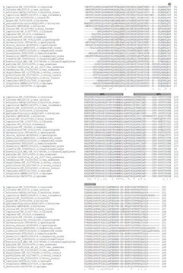
In spite of the evolutionary distance of the selected species, multiple alignment of the AIF-1 sequences from vertebrates, invertebrates, and choanoflagellates reveals highly conserved motifs and structural features (). In particular, AIF-1 protein sequence lengths span from 142 to 158 amino acids, with major sequence differences mainly discernable at the amino- and carboxy-terminal ends. Multiple sequence alignment shows the presence of 19 fully conserved amino acid residues (asterisks) and 32 positions exhibiting high conservation, thanks to the presence of amino acids with strongly similar properties (colons). The core of the proteins invariably contains two adjacent EF-hand (EFh) calcium-binding motifs, the second being less conserved than the first. Canonically, this type of domain consists of a 12-residue loop flanked on both sides by a 12-residue α-helical domain. Ca2+ binding induces a conformational change in the EFh motif, leading to the activation or inactivation of target proteins.
Figure 2. Multiple alignment among the sequences analyzed in as obtained by Clustal Omega (default parameters). EF-hand, calcium-binding motifs (Efh) are highlighted in grey. Asterisks (*) correspond to single, fully conserved residues. Colons (:) indicate conservation of residues sharing strongly similar properties (equivalent to scoring > 0.5 in the Gonnet PAM 250 matrix). Periods (.) indicate conservation of residues with weakly similar properties (equivalent to scoring ≤ 0.5 and > 0 in the Gonnet PAM 250 matrix).
2.1. Porifera
Porifera are the phylogenetically oldest still existent metazoans in which
AIF-1 has been identified
[28][29]. In these organisms, the protein presents a high sequence similarity with vertebrates. Kruse et al.
[28] observed that in the sponges
Suberites domuncula and
Geodia cydonium the expression of
AIF-1 mRNA was induced in cytokine-mediated allogeneic responses during wound repair. Interestingly, its expression does not occur in autografts, suggesting a possible function in immunocytes involved in alloimmune rejection
[28]. Cloning of
S. domuncula AIF-1 allows demonstration that the distribution of the six exon/intron borders is, with one exception, strictly conserved between sponge, human, and mouse genes. This also suggests a close evolutionary distance between these species
[29]. Moreover, in the same sponge it has been documented, both at tissue and in vitro level, that the expression of
AIF-1 and certain Tcf-like transcription factor genes is closely correlated with histoincompatibility reactions
[29]. Indeed,
AIF-1 expression is upregulated in transplants, especially in grafts deriving from different donors. According to Kruse et al.
[28], these data suggest that in sponges, in addition to an adaptive immunity, an effector system involving a cytokine-mediated activation of immunocytes occurs. Although we do not know which cells produce AIF-1 in sponges, it is possible to hypothesize that the ‘gray cells’, which are equivalent to the immune leukocytes within the vertebrates, are the main AIF-1 producers
[29].
2.2. Cnidaria
Apart from a gene sequence from
Nematostella vectensis present in GenBank (Acc. No. XP_001635454), data on AIF-1 in Cnidaria only derive from the sea anemone
Anemonia viridis, where an AIF-1 homologue was first identified and characterized
[31]. The predicted protein shows the common elements of AIF-1 family members, possessing the evolutionarily conserved EF-hand Ca
2+-binding motifs, the typical post-transcriptional modification sites, and a 3D structure that can be superimposed with human members of this family
[31]. Probably, in
A. viridis AIF-1 serves as a general protective factor under normal physiological conditions. However, after challenges with different stresses (i.e., biotic or physical challenge) a transcriptional activation can be observed, confirming the involvement of AIF-1 in the inflammatory response
[31]. In this anthozoan species, AIF-1 transcripts are detected in different tissues including the tentacles, oral disk, body wall, pharynx, and basal disk. Considering the basic diblastic organization of Cnidaria, these results evidence that in
A. viridis the expression pattern of AIF-1 could be distributed in the cells of both the ectoderm and endoderm layers
[31]. However, it is not clear at present whether the presence of this protein is limited to immunocytes, which possibly infiltrate the tissues.
2.3. Mollusca
Among invertebrates, due to their commercial interest, mollusks represent the most investigated species. Furthermore, frequently suffering from environmental bacterial infection, investigations in mollusks are prevalently targeted at the identification of genes modulated upon bacterial challenge. The first report of
AIF-1 in mollusks was in
Haliotis diversicolor hemocytes, where
AIF-1 was one of the 34 genes involved in different cellular pathways upregulated after bacterial challenge
[32]. Moreover, in this gastropod a high expression level of
AIF-1 mRNA was found in gills, suggesting that it could also have a significant contribution in the prevention of microbial infection
[33].
AIF-1 cDNA has also been cloned from Bivalves including oysters (
Pinctada martensii,
Crassostrea gigas, and
Crassostrea ariakensis)
[34][35][36], triangle sail mussel (
Hyriopsis cumingii)
[37], scallops (
Chlamys farreri)
[38], and clams (
Venerupis philippinarum)
[33], and also from gastropods such as abalones (
Haliotis discus discus)
[39]. Upregulation of
AIF-1 has been observed in some species after tissue injury such as tissue implant in
H. cumingii [37] or shell damage and mantle injury in
P. martensii [34]. In all these species, AIF-1 molecules were active in the host immune responses against pathogenic challenges. Indeed, in
V. philippinarum,
P. martensii, and
H. discus discus this gene was upregulated in hemocytes after infection by both Gram-positive and Gram-negative bacteria
[33][34][39], indicating its possible role in clearing pathogens soon after the infection. In support of this, there are also data showing the increased expression of
AIF-1 after LPS stimulation in
H. cumingii [37] and
C. ariakensis [36]. In the latter mollusk, using a recombinant AIF-1 protein, Xu et al.
[36] highlighted that AIF-1 acts in the regulation of some immune-related genes such as
LITAF,
MyD88, and
TGFβ. Interestingly, LITAF is an important transcription factor and is believed to regulate the expression of inflammatory-related factors IL-1α, TNFα, and IFN-γ in mammals
[40][41].
Studies on the role of AIF-1 in
H. cumingi evidenced a significant increase in the phagocytosis rate
[38]. This is not surprising since mollusk hemocytes, the main component of cellular immune responses in invertebrates, can be considered functionally analogous to vertebrate leukocytes, acting as macrophages and playing a crucial role in the recognition and removal of foreign materials
[42]. Recently, the functional role in hemocytes has been better pointed out by confocal imaging, which revealed that AIF-1 regulates phagocytosis via a functional interaction with filamentous actin
[43]. In hemocytes, AIF-1 appeared diffused in the cytoplasm and colocalized with F-actin bundles. After a bacterial challenge, a disruption of the AIF-1 and F-actin association and an increase in cell extension occurred. In all the investigated mollusks, the
AIF-1 gene resulted constitutively expressed in various tissues such as mantle, gill, hepatopancreas, muscle, and foot, with the highest level always being recorded in hemocytes
[33][39]. Some authors report the constitutive expression of
AIF-1 transcripts in a variety of unstimulated tissues. This is probably due to its involvement in various processes other than inflammation, pathogenic challenges, or tissue injury, which still need to be explored. However, although AIF-1 has been found in several organs, we cannot currently say whether it is expressed in cells other than hemocytes generally present in various tissues.
Gust et al.
[44] used the
AIF-1 gene as a marker to evaluate the immune effects of environmentally relevant concentrations of pharmaceutical mixtures on the pond snail
Lymnaea stagnalis. Results indicate that this factor, together with other immune and inflammatory markers, is modulated in the presence of drug mixtures and municipal effluent water in this gastropod. In particular, the
AIF-1 gene in
L. stagnalis is downregulated in response to antibiotic and psychiatric drug mixtures. This study demonstrates the interest of
AIF-1 as a potential biomarker for environmental studies on water chemical pollution.
2.4. Annelida
AIF-1 was initially characterized in annelids by Drago et al.
[45], who reported its presence, under the name of Iba1, in the CNS of the medicinal leech
Hirudo medicinalis. Iba1 is a largely recognized microglial marker in vertebrates, though it has not been detected in parenchymal brain microglia of zebrafish and birds
[46]. According to the authors, this work constitutes the first report of such a factor in the CNS of an invertebrate species
[45]. Like its vertebrate counterpart, the leech gene
Iba1/
AIF-1 is upregulated in nervous cells upon experimental injury or ATP stimulation. The predicted Iba1 protein shows an average identity of about 50% and 55% with AIF-1 proteins described in vertebrates and invertebrates, respectively. Immunohistochemistry analyses demonstrated its presence in activated microglia accumulated at the injury site of connective fibers and in those surrounding neuron cell bodies. Results from Schorn et al.
[47] established the constitutive presence of the Iba1/AIF-1 protein in CD68+ and CD45+ macrophage-like cells spread in leech body wall tissues. The number of AIF-1 immunopositive cells strongly increases upon bacterial challenge. In addition, the injection of recombinant AIF-1 in leeches induces massive angiogenesis and, similarly to AIF-1 in vertebrates, promotes macrophage-like cell recruitment at the injured site. Recent works from the same team
[48][49] demonstrated that RNASET2, a protein belonging to the T2 ribonuclease family, and AIF-1 are released from the same immunocompetent cells in the leech
H. verbana8. Both factors would contribute to the recruitment of immune cells (granulocytes and macrophages) upon LPS injection or wound healing, resulting in the activation of an effective response against pathogen infection. AIF-1 has also been identified in
Hirudo telocytes
[50], special resident cells involved in the immune-surveillance system of the leech body wall. Like in vertebrates, these cells also play a role in regulating immune and neuroendocrine functions in leeches. All together, these data indicate the involvement of AIF-1 in immune cell activation and migration as well as in regulating the inflammatory response in leeches.
2.5. Echinodermata
Echinoderms represent the most developed invertebrates and the bridge leading to the primitive chordates, cephalochordates, and urochordates, in which many autologous genes and functions from their ancestors can be found.
The evolutionary position of echinoderms among the Deuterostomia, the same evolutionary branch where vertebrates are found, represents one of the reasons that makes these animals attractive as upcoming model systems. Echinoderms, besides being an important source of food and medicine for humans, provide important clues to understand immune functions that are common with vertebrates. Firstly, Elie Metchnikoff, by introducing the comparative approach to immunology
[51], postulated the inflammation concept applicable to mammals and other animals with closed circulatory systems. Metchnikoff’s study in this field started from the observation of phagocyte recruitment around foreign material in the larvae of an echinoderm (sea star)
[52].
Only many years later were the immune and non-self recognition capabilities established in these organisms
[53][54][55][56] when molecular approaches were developed to evidence novel immune effectors.
The first report on the presence of
AIF-1 in echinoderms
[57] was based on the EST analysis of genes upregulated in coelomocytes in response to LPS challenge. Indeed, Nair et al.
[57] identified one gene, named
Sp1086, matching allograft inflammatory factor-1 in the purple sea urchin,
Strongylocentrotus purpuratus.
Within the Echinodermata phylum, the sea urchin represents an excellent model organism for studies on inflammation, including those on the expression and regulation of
AIF-1. Indeed, the Antarctic sea urchin
Sterechinus neumayeri [58] and the common sea urchin
Paracentrotus lividus [59][60] have furnished intriguing information about the association of
AIF-1 with the immune response. AIF-1 has been identified in coelomocytes of
S. neumayeri, where an increase in expression during the first phase of the immune response to a bacterial challenge was evidenced. Interestingly, with
S. neumayeri being a sea urchin living in circumpolar waters, the protein primary structure presents some molecular adaptations to cold
[58], and this report indicates that
AIF-1 can participate in the inflammatory response in extremely cold environments.
Further data have been derived from investigation in the common sea urchin
P. lividus, where the molecular identification and functional characterization of
AIF-1 have been recently reported
[61]. In this work, the authors found a significant increase in
AIF-1 expression, at both the mRNA and protein level, in coelomocytes after Gram+ bacterial challenge. In addition, immunocytochemical analysis conducted on different coelomocyte populations revealed the presence of the AIF-1 protein in the perinuclear cytoplasmic zone of amoebocytes and inside red sphaerula cell granules. With these cells being involved in the inflammatory reaction, it is possible to support that AIF-1 plays a crucial role in the defense processes within echinoderms. More recently, information on the
AIF-1 gene in
P. lividus has been enriched by the work of Chiaramonte et al.
[60], who reported its modulation following LPS challenge and the bioinformatically characterized protein structure.
In the sea cucumber
Apostichopus japonicus, the full-length cDNA of
AIF-1 has been cloned
[61]. Like the sea urchin, in this organism a significant increase in the expression levels of
AIF-1 transcripts has also been detected in coelomocytes after bacterial challenge and papilla injury. Based on these results, the authors supported the idea that AIF-1 is involved in acute inflammatory response. Furthermore, data reported the constitutive expression of
AIF-1 in all the tested tissues, including body wall, intestine, respiratory tree, tube feet, and longitudinal muscle.
 +1 point